Copyright
©The Author(s) 2016.
World J Stem Cells. Dec 26, 2016; 8(12): 399-427
Published online Dec 26, 2016. doi: 10.4252/wjsc.v8.i12.399
Published online Dec 26, 2016. doi: 10.4252/wjsc.v8.i12.399
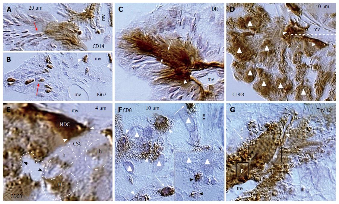
Figure 20 Ovarian cancer augmentation by the immune/morphostatic system.
A: Association of CD14+ MDC (arrowhead) with cancer microvasculature (mv) results in nuclear and cytoplasmic CD14 expression by cancer/MDC hybrids (white arrow) and nuclear CD14 expression in more distant cancer cells (red arrow); B: Ki67 expression by divided perivascular (arrowheads) and postmitotic malignant cells (arrow); C: HLA DR+ expression by perivascular MDCs (arrowheads) and adjacent cancer cells (arrows); D: CD68 MDCs associate with microvasculature (arrowheads) and most malignant cells express CD68 (triangles); E: Detail from panel D shows an apposition (white arrowheads) of perivascular CD68 MDC with unstained cancer stem cell (CSC). The fresh cancer/normal cell hybrids (h) show partial CD68 expression and more advanced hybrids (H) are more distinctly stained. Dividing cancer stem cell (dotted line) is unstained; F: CD8 T cells (TC) evade (arrows) from cancer microvasculature (mv) and exhibit increase in size (arrowheads) among cancer cells (triangles). Inset shows regressing TC (arrowheads) among cancer cells; G: Cancer microvasculature (mv) is accompanied by hyperactive pericytes (p) producing Thy-1+ vesicles (black arrowhead) releasing their growth promoting substances among adjacent cancer cells and collapsing into the Thy-1+ spikes (arrow). The microvasculature produces new endothelial cells (e) accompanied by new Thy-1+ pericytes (white arrowhead). Adjusted from[21]: ©Antonin Bukovsky.
- Citation: Bukovsky A. Involvement of blood mononuclear cells in the infertility, age-associated diseases and cancer treatment. World J Stem Cells 2016; 8(12): 399-427
- URL: https://www.wjgnet.com/1948-0210/full/v8/i12/399.htm
- DOI: https://dx.doi.org/10.4252/wjsc.v8.i12.399


