Copyright
©The Author(s) 2016.
World J Stem Cells. Dec 26, 2016; 8(12): 399-427
Published online Dec 26, 2016. doi: 10.4252/wjsc.v8.i12.399
Published online Dec 26, 2016. doi: 10.4252/wjsc.v8.i12.399
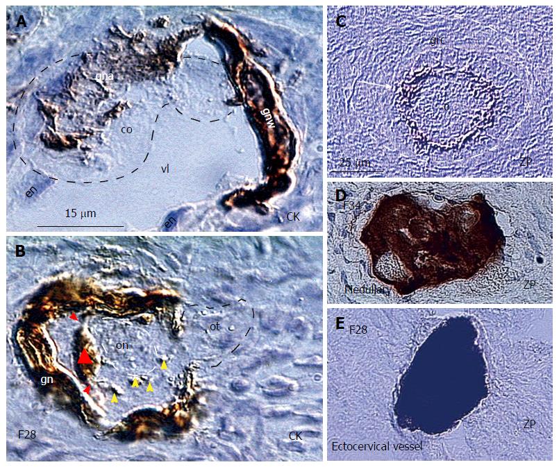
Figure 12 Granulosa cells are essential for the in vivo survival of the newly formed oocytes.
A: The small circulating oocyte (co) is captured in the lower ovarian cortex by CK+ granulosa nest arm (gna) in the vein lumen (vl) lined by endothelial cells (en) and granulosa nest wall (gnw); B: The CK+ primary Balbiani body (triangle) adjacent to the oocyte nucleus (on) is formed by granulosa cell extensions (red arrowheads) and disperses its particles (yellow arrowheads) to the freshly captured oocyte, which still exhibits the oocyte tail (ot) outside of the nest; C: Intact oocyte (o) covered by ZP+ membrane (arrow) and granulosa cells (grc) in the primary follicle (dashed line); D: The vein in ovarian medulla in the identical ovary contains regressing oocyte with marked cytoplasmic ZP staining; E: Regressing oocyte with strong cytoplasmic ZP expression found in the ectocervical vein in the women showing follicular renewal in A and B panels. Panels A and B adjusted from[43]: © Antonin Bukovsky; C-E from[172], with a permission: ©Elsevier/North-Holland Biomedical Press. The panels A, B, and E are from the identical ovary of 28-year-old women presented in the Figure 10.
- Citation: Bukovsky A. Involvement of blood mononuclear cells in the infertility, age-associated diseases and cancer treatment. World J Stem Cells 2016; 8(12): 399-427
- URL: https://www.wjgnet.com/1948-0210/full/v8/i12/399.htm
- DOI: https://dx.doi.org/10.4252/wjsc.v8.i12.399


