Copyright
©The Author(s) 2016.
World J Stem Cells. Dec 26, 2016; 8(12): 399-427
Published online Dec 26, 2016. doi: 10.4252/wjsc.v8.i12.399
Published online Dec 26, 2016. doi: 10.4252/wjsc.v8.i12.399
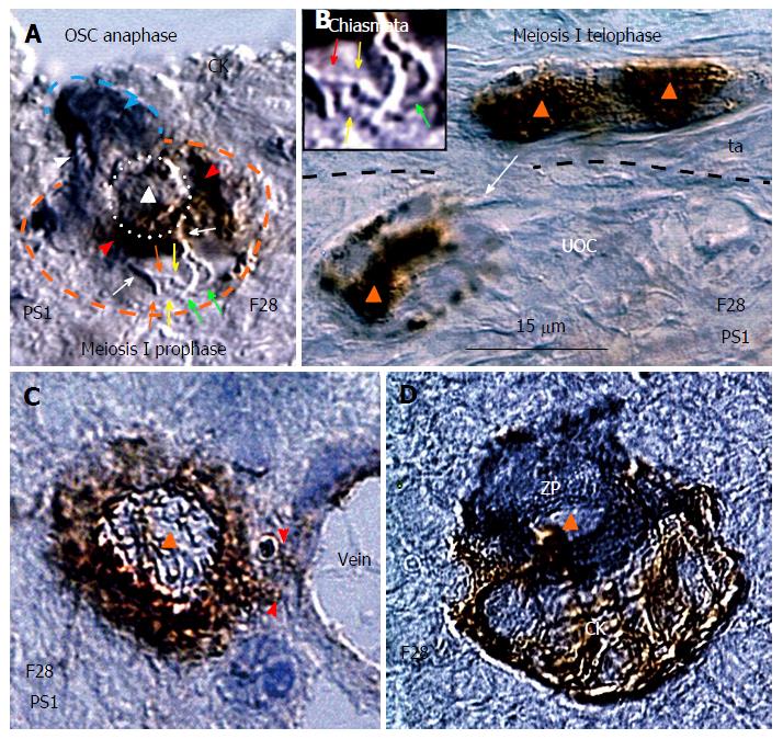
Figure 10 Meiotic events during midfollicular phase are followed by follicular renewal.
A: Cytokeratin stained ovarian stem cell (OSC) (blue arrowhead) moves its chromosomes (white arrowhead) during the OSC anaphase to the OSC end. The germ cell expressing PS1 meiotic protein (red arrowhead) exhibits meiosis I prophase with chromosome (white arrows) duplication (red and green arrows) and crossover of sister chromatids (yellow arrows). The triangle indicates putative suicidal T cell (see Figure 9B) inducing expression of PS1 in the emerging germ cel; B: Marked nuclear expression of PS1 accompanies germ cell telophase of meiosis I in the tunica albuginea (ta). Arrow indicates migrating postmeiotic germ cell. Inset shows a detail of interacting chromosomes (red and green arrows) from panel A; C: The germ cell entering (arrowheads) the cortical vein exhibits cytoplasmic but not nuclear (triangle) PS1 staining; D: Association of zona pellucida+ small oocyte with CK+ granulosa cell nest during the new primary follicle formation in the lower ovarian cortex[43]. All panels are from the identical ovary during midfollicular phase of the 28-year-old women.
- Citation: Bukovsky A. Involvement of blood mononuclear cells in the infertility, age-associated diseases and cancer treatment. World J Stem Cells 2016; 8(12): 399-427
- URL: https://www.wjgnet.com/1948-0210/full/v8/i12/399.htm
- DOI: https://dx.doi.org/10.4252/wjsc.v8.i12.399


