Copyright
©The Author(s) 2016.
World J Stem Cells. Dec 26, 2016; 8(12): 399-427
Published online Dec 26, 2016. doi: 10.4252/wjsc.v8.i12.399
Published online Dec 26, 2016. doi: 10.4252/wjsc.v8.i12.399
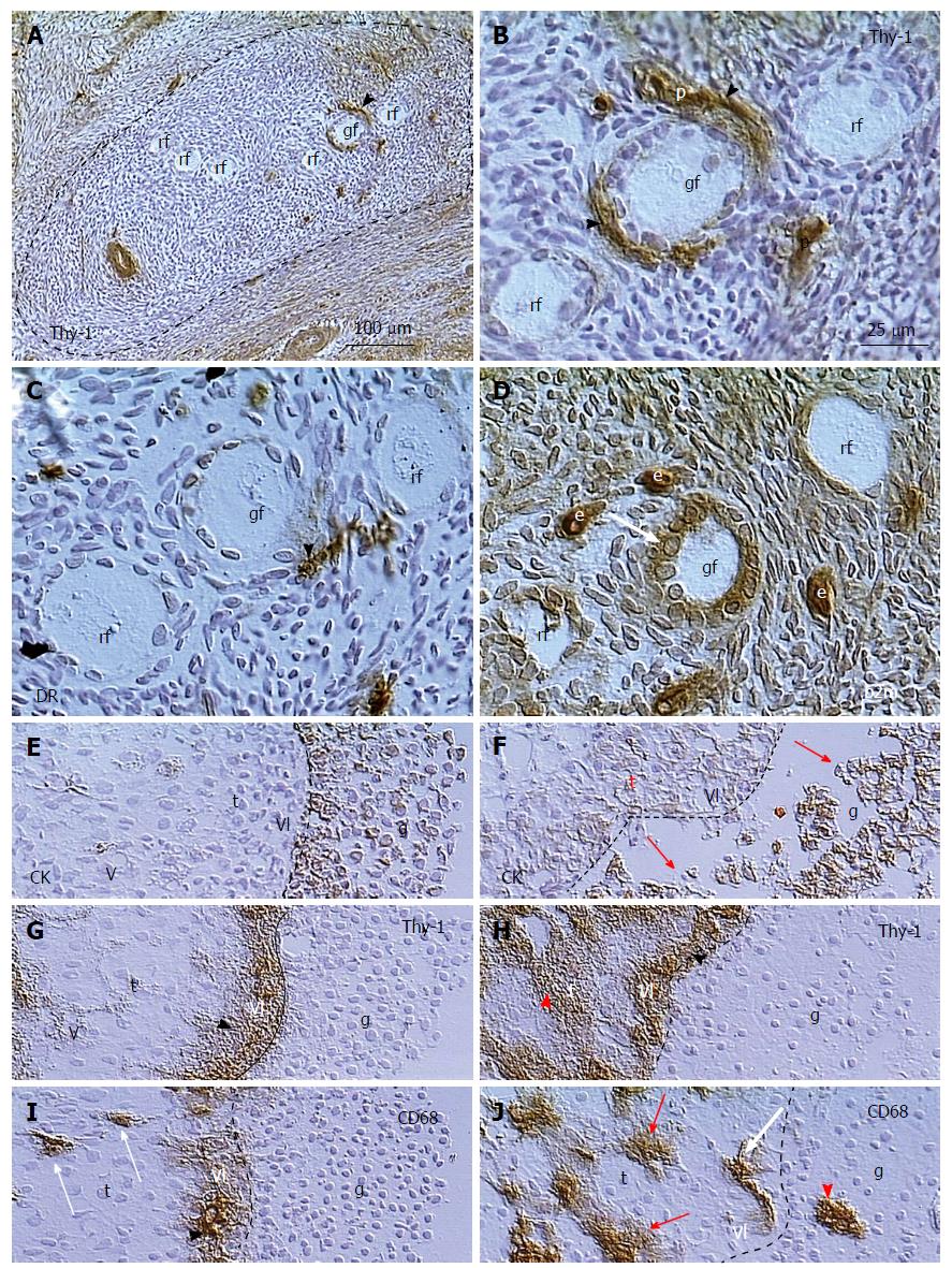
Figure 7 Follicular selection.
A: Resting primary follicles (rf) are present in ovarian area without Thy-1 expression by stromal cells, where growing primary follicle (gf) receives Thy-1 supply from vascular pericytes (arrowhead); B: Detail from (A) shows Thy-1 supply (arrowheads) from vascular pericytes (p) for the growing primary follicle; C: Parallel section showing association of DR+ activated MDC (arrowhead) with the growing but not resting primary follicles; D: Growing primary follicle exhibits large granulosa cells with strong expression of beta2m; E: Dominant preovulatory follicle granulosa cells (g) with expression of cytokeratin (CK) are attached to the follicular basement membrane (dashed line). Note lack of staining in vascular lamina propria (vl) and theca interna cells (t); F: Regressing large antral follicle in the same ovary exhibits detachment of granulosa cells (arrows) and CK expression in the vascular lamina and thecal cells; G: Thy-1 expression in vascular lamina (arrowhead) but no staining of theca interna in dominant follicle; H: Regressing follicle with Thy-1 staining in both, vascular lamina propria and theca interna (red arrowhead); I: CD68 is released from MDCs in the vascular lamina (arrowhead) of the dominant follicle, but not from thecal MDCs (arrows), and no MDCs are present among the granulosa cells; J: In the regressing follicle the MDCs release CD68 in theca interna (red arrows), and invade among granulosa cells (arrowhead)[171].
- Citation: Bukovsky A. Involvement of blood mononuclear cells in the infertility, age-associated diseases and cancer treatment. World J Stem Cells 2016; 8(12): 399-427
- URL: https://www.wjgnet.com/1948-0210/full/v8/i12/399.htm
- DOI: https://dx.doi.org/10.4252/wjsc.v8.i12.399


