Copyright
©The Author(s) 2016.
World J Stem Cells. Dec 26, 2016; 8(12): 399-427
Published online Dec 26, 2016. doi: 10.4252/wjsc.v8.i12.399
Published online Dec 26, 2016. doi: 10.4252/wjsc.v8.i12.399
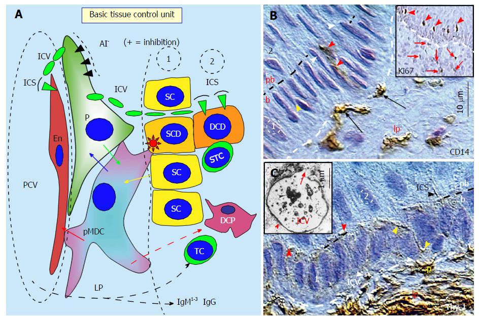
Figure 1 The basic tissue control unit and early cellular differentiation.
A: The tissue control unit (TCU) is associated with postcapillary venules (PCV). It consists of CD14+ primitive MDCs (pMDCs), pericytes (P) accompanying PCV, and autonomic innervation (AI). The TCU influences properties of Endothelial cells (En) and an involvement of other components of the TCS regulating the differentiation of tissue stem cells into the tissue-specific functional stage by the influence of DCP dendritic cell precursors (DCP), and eventually by T cells (TC), dendritic cells, and immunoglobulins (IgM1-3 and IgG). The pMDCs physically interact with adjacent En (red arrow) and receive requests (yellow arrow) to regenerate from tissue stem cells (SC) when required. The pMDCs communicate with pericytes (blue arrow), and if the pericytes are not blocked by AI, the positive signal (green arrow) is provided to pMDC to stimulate stem cell division. The asymmetric division is initiated by pMDC (red asterisk) and accompanied by a suicidal T cell (STC). It gives a rise to the stem cell daughter (SCD) and differentiating cell daughter (DCD). The pericytes provide by Thy-1+ intercellular vesicles (ICV) growth factors and cytokines to the endothelial and tissue cells. After release of ICV content (green arrowheads), the vesicles collapse into intercellular spikes (ICS); B: CD14 MDCs (arrows) in lamina propria (lp) migrate to basal layer (b) of the stratified epithelium, interact with basal stem cells (yellow arrowhead), and migrate to the parabasal layer (red arrowheads). White arrowheads indicate basal epithelial cells mowing to the parabasal (pb) layer. Inset shows Ki67+ postmitotic parabasal epithelial cells (arrowheads) represented by differentiating stem cell daughters, and postmitotic stromal cells in the lamina propria (arrows); C: Thy-1 P in the lamina propria produce ICV (white arrowheads) migrating (yellow arrowheads) toward postmitotic parabasal cells (red arrowheads) where they release their content and collapse into ICS (black arrowhead). Inset shows transmission electron microscopy of Thy-1 immunolabeling of a pericyte-derived ICV releasing its content (arrow)[1].
- Citation: Bukovsky A. Involvement of blood mononuclear cells in the infertility, age-associated diseases and cancer treatment. World J Stem Cells 2016; 8(12): 399-427
- URL: https://www.wjgnet.com/1948-0210/full/v8/i12/399.htm
- DOI: https://dx.doi.org/10.4252/wjsc.v8.i12.399


