Copyright
©The Author(s) 2015.
World J Stem Cells. Aug 26, 2015; 7(7): 1047-1053
Published online Aug 26, 2015. doi: 10.4252/wjsc.v7.i7.1047
Published online Aug 26, 2015. doi: 10.4252/wjsc.v7.i7.1047
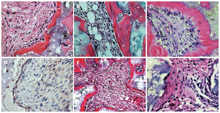
Figure 4 Generation of cementum-like and PDL-like structures in vivo by PDLSCs.
A: After 8 wk of transplantation, PDLSCs differentiated into cementoblast-like cells (arrows) that formed a cementum-like structure (C) on the surface of the hydroxyapatite tricalcium phosphate (HA) carrier; cementocyte-like cells (arrowhead) and PDL-like tissue (PDL) were also generated; B: BMSSC transplant was used as control to show the formation of a bone/marrow structure containing osteoblasts (arrows), osteocytes (arrowhead), and elements of bone (B) and haemopoietic marrow (HP); C: DPSC transplant was also used as a control to show a dentin/pulp-like structure containing odontoblasts (arrows) and dentin like (D) and pulp-like (Pulp) tissue; D: Immunohistochemical staining showed that PDLSCs generated cementum-like structure (C) and differentiated into cementoblast-like cells (arrows) and cementocyte-like cells (arrowhead) that stained positive for human-specific mitochondria antibody. Part of the PDL-like tissue (PDL) also stained positive for human specific mitochondria antibody (within dashed line); E: Of 13 selected strains of single-colony derived PDLSC, only eight (61%) generated cementum/PDL-like structures in vivo as shown at lower magnification (approximately 20). New cementum-like structure (C) formed adjacent to the surfaces of the carrier (HA) and associated with PDL-like tissue (PDL); F: The othser five strains did not generate mineralised or PDL-like tissues in vivo[38].
- Citation: Aly LAA. Stem cells: Sources, and regenerative therapies in dental research and practice. World J Stem Cells 2015; 7(7): 1047-1053
- URL: https://www.wjgnet.com/1948-0210/full/v7/i7/1047.htm
- DOI: https://dx.doi.org/10.4252/wjsc.v7.i7.1047


