Copyright
©The Author(s) 2015.
World J Stem Cells. May 26, 2015; 7(4): 728-744
Published online May 26, 2015. doi: 10.4252/wjsc.v7.i4.728
Published online May 26, 2015. doi: 10.4252/wjsc.v7.i4.728
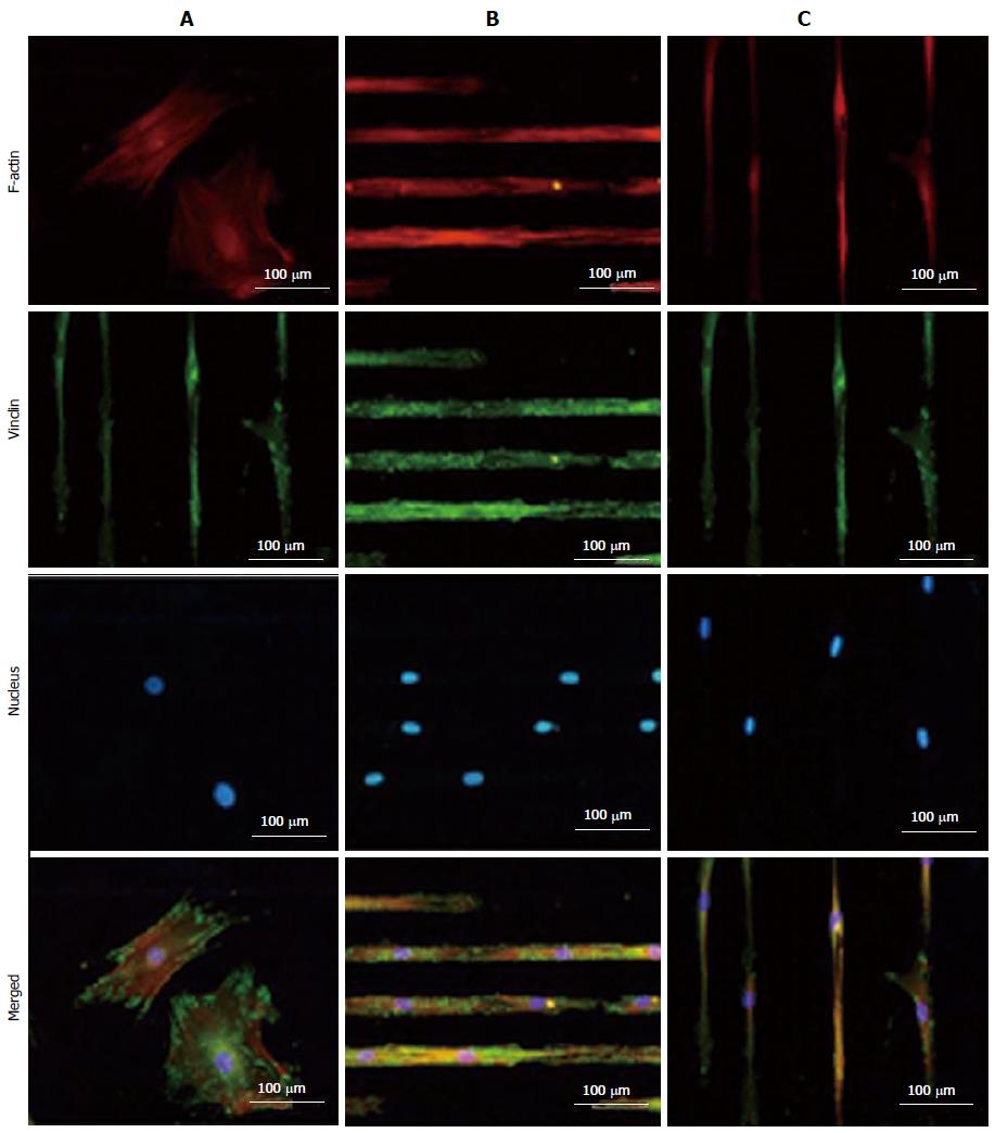
Figure 3 TRITC-Phalloidin labeled F-actin (red), AlexaFluor 488 labeled vinculin (green), diamidino-2-phenylindole nuclear staining (blue) and overlaid fluorescent image of immuno-stained cellular components (merged) for the unpatterned (A) and patterned human mesenchymal stem cells (B and C).
Samples were cultured in Dulbecco's Modified Eagle's Medium supplemented with 10% fetal bovine serum and 1% antibiotic/antimycotic solution for 4 d before they were fixed and stained. All images were taken with a 20 × objective lens. (Scale bar = 100 μm) (For interpretation of the references to colour in this figure legend, the reader is referred to the web version of this article.). Reproduced with permission from Tay et al[86].
- Citation: Ghasemi-Mobarakeh L, Prabhakaran MP, Tian L, Shamirzaei-Jeshvaghani E, Dehghani L, Ramakrishna S. Structural properties of scaffolds: Crucial parameters towards stem cells differentiation. World J Stem Cells 2015; 7(4): 728-744
- URL: https://www.wjgnet.com/1948-0210/full/v7/i4/728.htm
- DOI: https://dx.doi.org/10.4252/wjsc.v7.i4.728


