Copyright
©2014 Baishideng Publishing Group Co.
World J Stem Cells. Apr 26, 2014; 6(2): 134-143
Published online Apr 26, 2014. doi: 10.4252/wjsc.v6.i2.134
Published online Apr 26, 2014. doi: 10.4252/wjsc.v6.i2.134
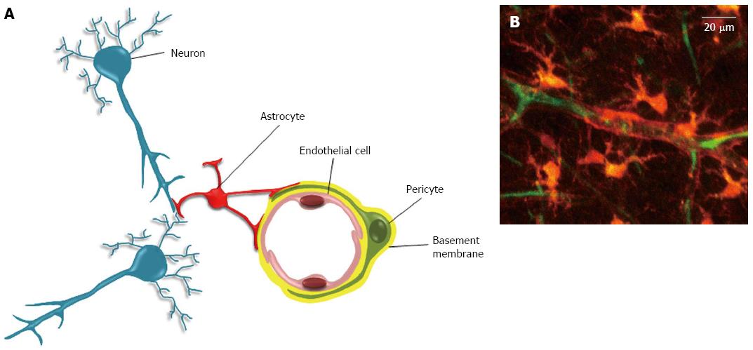
Figure 2 The neurovascular unit.
A: The neurovascular unit. In the neurovascular unit, pericytes are located on the abluminal side of endothelial cells (EC). Both cells are ensheathed by the basement membrane (BM). The covering of EC by pericytes is incomplete, and interruptions in BM can allow direct contacts between pericyte and EC. These contacts occur through peg and socket structures, and adherent and gap junctions (not shown)[59]. The abluminal side of the basement membrane is also contacted by astrocytes endfeet. In addition to these cells, the neurovascular unit also includes neurons, and microglial cells (not shown); B: Two-photon microscopy of a neurovascular unit. Following injection in the rat tail vein, the sulforhodamine-B dye crosses the blood brain barrier and stains astrocytes and pericytes in orange (reproduced from[115]). The blood plasma is shown in green after iv injection of FITC-dextran (Mw 70 kDa). Neurons, endothelial and microglial cells are not shown here.
- Citation: Appaix F, Nissou MF, Sanden BVD, Dreyfus M, Berger F, Issartel JP, Wion D. Brain mesenchymal stem cells: The other stem cells of the brain? World J Stem Cells 2014; 6(2): 134-143
- URL: https://www.wjgnet.com/1948-0210/full/v6/i2/134.htm
- DOI: https://dx.doi.org/10.4252/wjsc.v6.i2.134


