Copyright
©2013 Baishideng Publishing Group Co.
World J Stem Cells. Jan 26, 2013; 5(1): 26-33
Published online Jan 26, 2013. doi: 10.4252/wjsc.v5.i1.26
Published online Jan 26, 2013. doi: 10.4252/wjsc.v5.i1.26
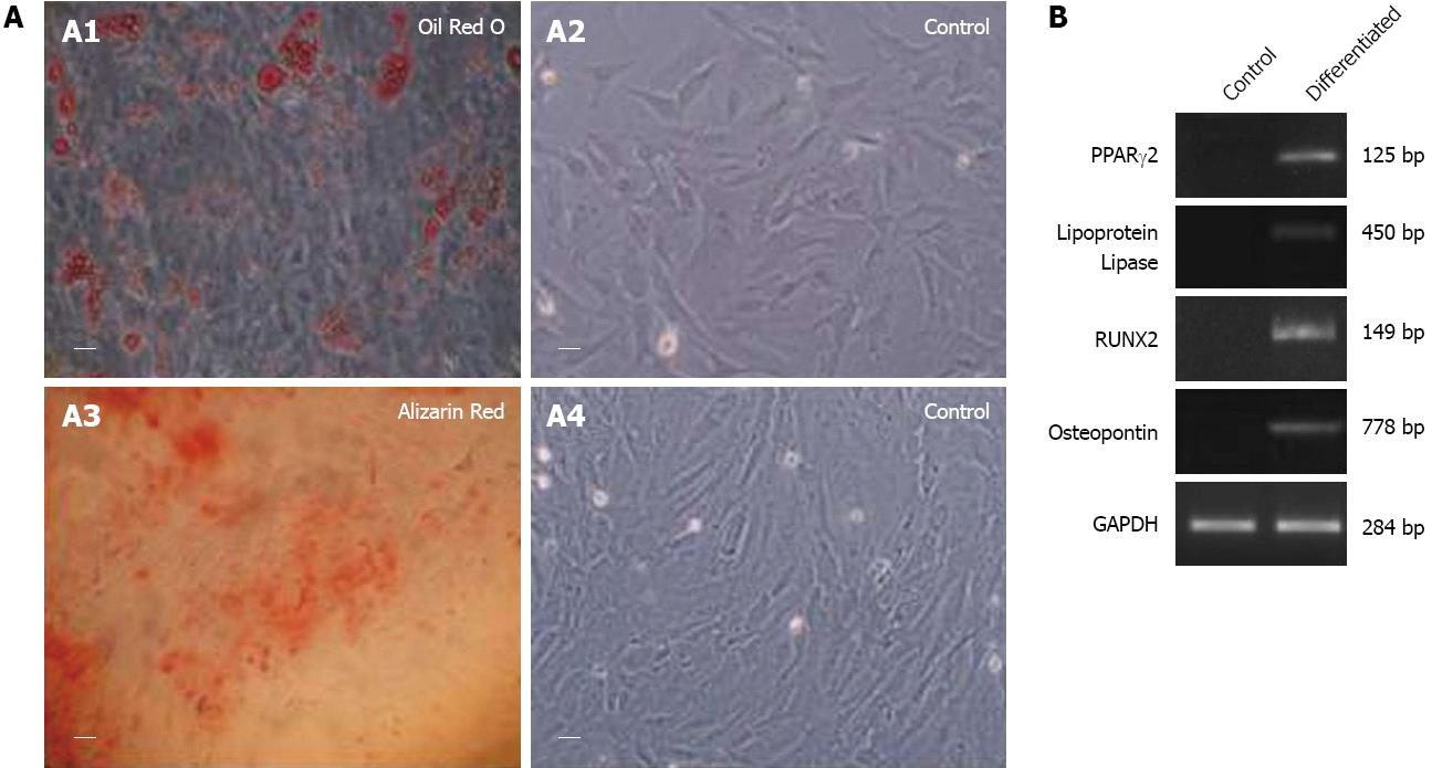
Figure 3 Representative photomicrographs (A) and representative reverse-transcription polymerase chain reaction gel photomicrographs (B).
A: Representative photomicrographs (40 ×, 20 μm) showing differentiation of rat fetal cardiac mesenchymal stem cells into osteocytes (A1: differentiated cells positive for Alizarin Red stain and A2: control cells negative for Alizarin Red stain) and adipocytes (A3: differentiated cells positive for Oil Red O stain and A4: control cells negative for Oil Red O stain); B: Representative reverse-transcription polymerase chain reaction gel photomicrographs showing expression of lipoprotein lipase and PPARγ2 by adipocytes and osteopontin and RUNX2 by osteocytes cells, induced from rat fetal cardiac mesenchymal stem cells. Control cells not treated with induction medium did not show expression of above markers.
- Citation: Srikanth GVN, Tripathy NK, Nityanand S. Fetal cardiac mesenchymal stem cells express embryonal markers and exhibit differentiation into cells of all three germ layers. World J Stem Cells 2013; 5(1): 26-33
- URL: https://www.wjgnet.com/1948-0210/full/v5/i1/26.htm
- DOI: https://dx.doi.org/10.4252/wjsc.v5.i1.26


