Copyright
©2013 Baishideng Publishing Group Co.
World J Stem Cells. Jan 26, 2013; 5(1): 26-33
Published online Jan 26, 2013. doi: 10.4252/wjsc.v5.i1.26
Published online Jan 26, 2013. doi: 10.4252/wjsc.v5.i1.26
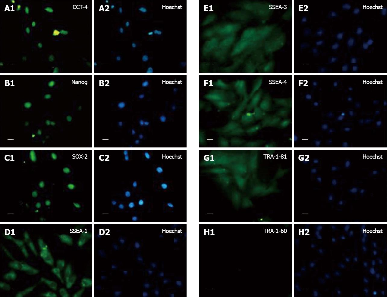
Figure 2 Representative immunocytochemistry photomicrographs (40 ×, 20 μm) of rat fetal cardiac mesenchymal stem cells showing expression.
A: OCT-4 (A1: OCT-4 and A2: Hoechst dye); B: Nanog (B1: Nanog and B2: Hoechst dye); C: SOX-2 (C1: SOX-2 and C2: Hoechst dye); D: SSEA-1 (D1: SSEA-1 and D2: Hoechst dye); E: SSEA-3 (E1: SSEA-3 and E2: Hoechst dye); F: SSEA-4 (F1: SSEA-4 and F2: Hoechst dye); G: TRA 1-81 (G1: TRA 1-81 and G2: Hoechst dye); H: TRA1-60 (H1:TRA 1-60 and H2: Hoechst dye).
- Citation: Srikanth GVN, Tripathy NK, Nityanand S. Fetal cardiac mesenchymal stem cells express embryonal markers and exhibit differentiation into cells of all three germ layers. World J Stem Cells 2013; 5(1): 26-33
- URL: https://www.wjgnet.com/1948-0210/full/v5/i1/26.htm
- DOI: https://dx.doi.org/10.4252/wjsc.v5.i1.26


