
















©The Author(s) 2025.
World J Stem Cells. Aug 26, 2025; 17(8): 107124
Published online Aug 26, 2025. doi: 10.4252/wjsc.v17.i8.107124
Published online Aug 26, 2025. doi: 10.4252/wjsc.v17.i8.107124
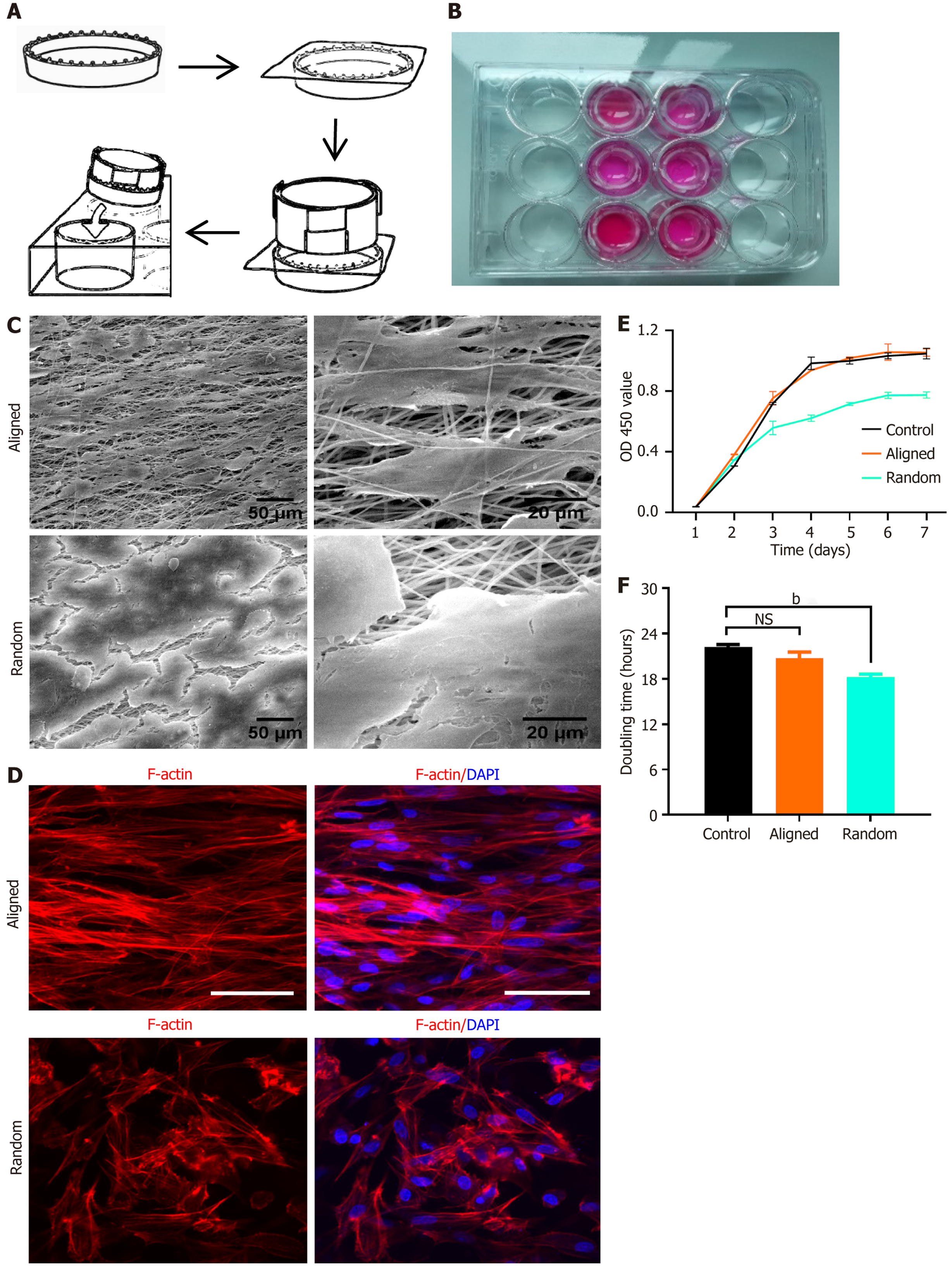
Figure 3 Bone marrow-derived mesenchymal stem cell seeding, morphology, and viability on aligned and random nanofiber scaffolds.
A: Schematic illustration of bone marrow-derived mesenchymal stem cell (BMSC) seeding on aligned and random nanofiber scaffolds placed in culture wells; B: Image of culture wells containing scaffolds with uniform BMSC seeding; C: Scanning electron microscopy images of aligned (top) and random (bottom) nanofiber scaffolds, highlighting organized orientation in aligned fibers and disordered structure in random fibers. Scale bars: 50 μm and 20 μm; D: Immunofluorescent staining for F-actin (red) and nuclei (4’,6-diamidino-2-phenylindole, blue) illustrating BMSC morphology and cytoskeletal arrangement on aligned vs random scaffolds. BMSCs on aligned scaffolds show an elongated, aligned cytoskeletal organization, while those on random scaffolds exhibit a more dispersed morphology; E: Cell viability assay using CCK-8 over 7 days, showing OD values at 450 nm for BMSCs on aligned, random, and control surfaces; F: Doubling time analysis of BMSCs on each substrate, with aligned scaffolds supporting faster proliferation than the random and control conditions. bP < 0.01, NS: Not significant. DAPI: 4’,6-diamidino-2-phenylindole.
- Citation: Yang CW, Zhang YQ, Chang H, Gao R, Chen D, Yao H. Aligned nanofiber scaffolds combined with cyclic stretch facilitate mesenchymal stem cell differentiation for ligament engineering. World J Stem Cells 2025; 17(8): 107124
- URL: https://www.wjgnet.com/1948-0210/full/v17/i8/107124.htm
- DOI: https://dx.doi.org/10.4252/wjsc.v17.i8.107124