Copyright
©The Author(s) 2025.
World J Stem Cells. Aug 26, 2025; 17(8): 107124
Published online Aug 26, 2025. doi: 10.4252/wjsc.v17.i8.107124
Published online Aug 26, 2025. doi: 10.4252/wjsc.v17.i8.107124
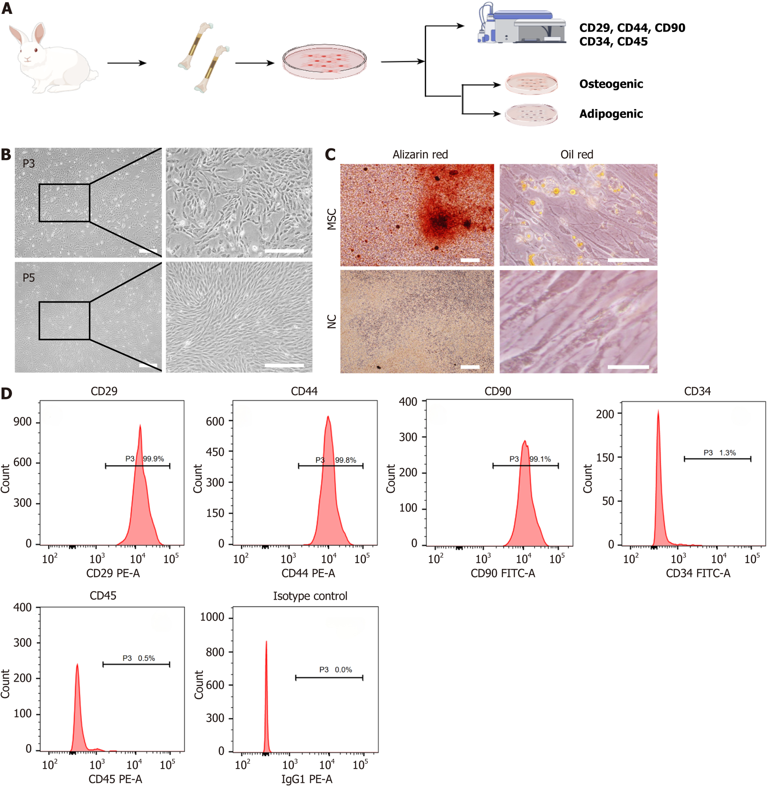
Figure 2 Isolation and characterization of rabbit mesenchymal stem cells.
A: Schematic representation of the mesenchymal stem cell (MSC) isolation and culture process from rabbit bone tissue, followed by phenotypic characterization through surface markers and differentiation assays; B: Phase-contrast images of MSCs at passage 3 and passage 5 showing spindle-shaped morphology. Scale bar: 100 μm; C: Differentiation assays demonstrating osteogenic and adipogenic potential, with positive Alizarin Red staining for osteogenesis and Oil Red O staining for adipogenesis in the MSC group; the negative control shows no staining. Scale bar: 100 μm; D: Flow cytometry analysis of surface markers, showing high expression of CD29, CD44, and CD90, and low expression of CD34 and CD45, confirming the MSC phenotype. P3: Passage 3; P5: Passage 5; MSC: Mesenchymal stem cell; NC: Negative control.
- Citation: Yang CW, Zhang YQ, Chang H, Gao R, Chen D, Yao H. Aligned nanofiber scaffolds combined with cyclic stretch facilitate mesenchymal stem cell differentiation for ligament engineering. World J Stem Cells 2025; 17(8): 107124
- URL: https://www.wjgnet.com/1948-0210/full/v17/i8/107124.htm
- DOI: https://dx.doi.org/10.4252/wjsc.v17.i8.107124


