Copyright
©The Author(s) 2025.
World J Stem Cells. May 26, 2025; 17(5): 101898
Published online May 26, 2025. doi: 10.4252/wjsc.v17.i5.101898
Published online May 26, 2025. doi: 10.4252/wjsc.v17.i5.101898
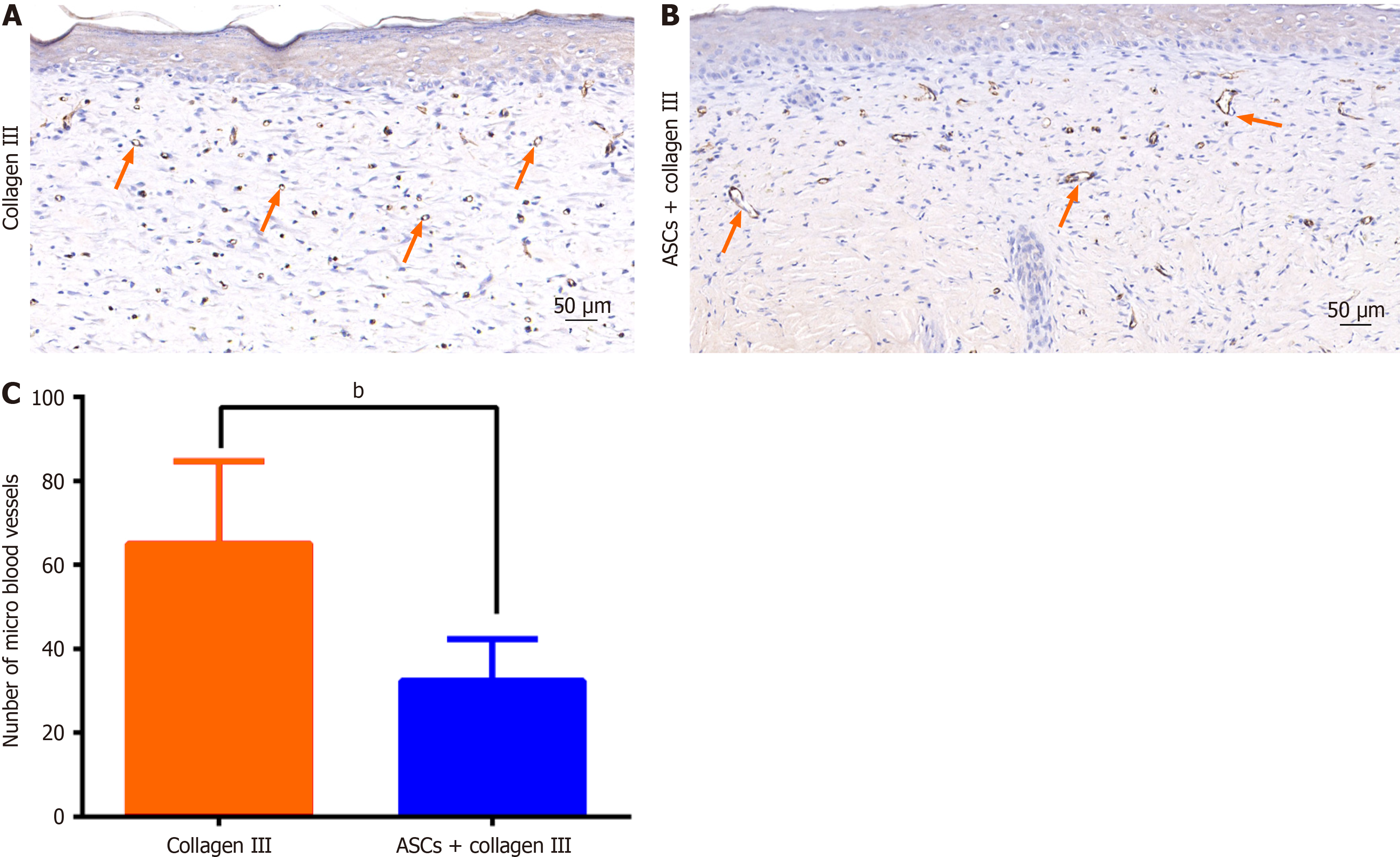
Figure 3 Immunohistochemical staining against CD31.
A: There were more micro-blood vessels in the collagen III group; B: Larger blood vessels were found in the treatment group. The orange arrows indicate some key areas of interest within the figure; C: The average number of micro blood vessels of the dermal layer was significantly different (n = 9, P < 0.0001). bP < 0.01. ASCs: Adipose-derived stem cells.
- Citation: Zhou XL, Wu B, Xie ZJ, Li HD. Collagen III combined with autologous adipose-derived mesenchymal stem cells accelerates burn wound healing in a rat model. World J Stem Cells 2025; 17(5): 101898
- URL: https://www.wjgnet.com/1948-0210/full/v17/i5/101898.htm
- DOI: https://dx.doi.org/10.4252/wjsc.v17.i5.101898


