Copyright
©The Author(s) 2022.
World J Stem Cells. Sep 26, 2022; 14(9): 700-713
Published online Sep 26, 2022. doi: 10.4252/wjsc.v14.i9.700
Published online Sep 26, 2022. doi: 10.4252/wjsc.v14.i9.700
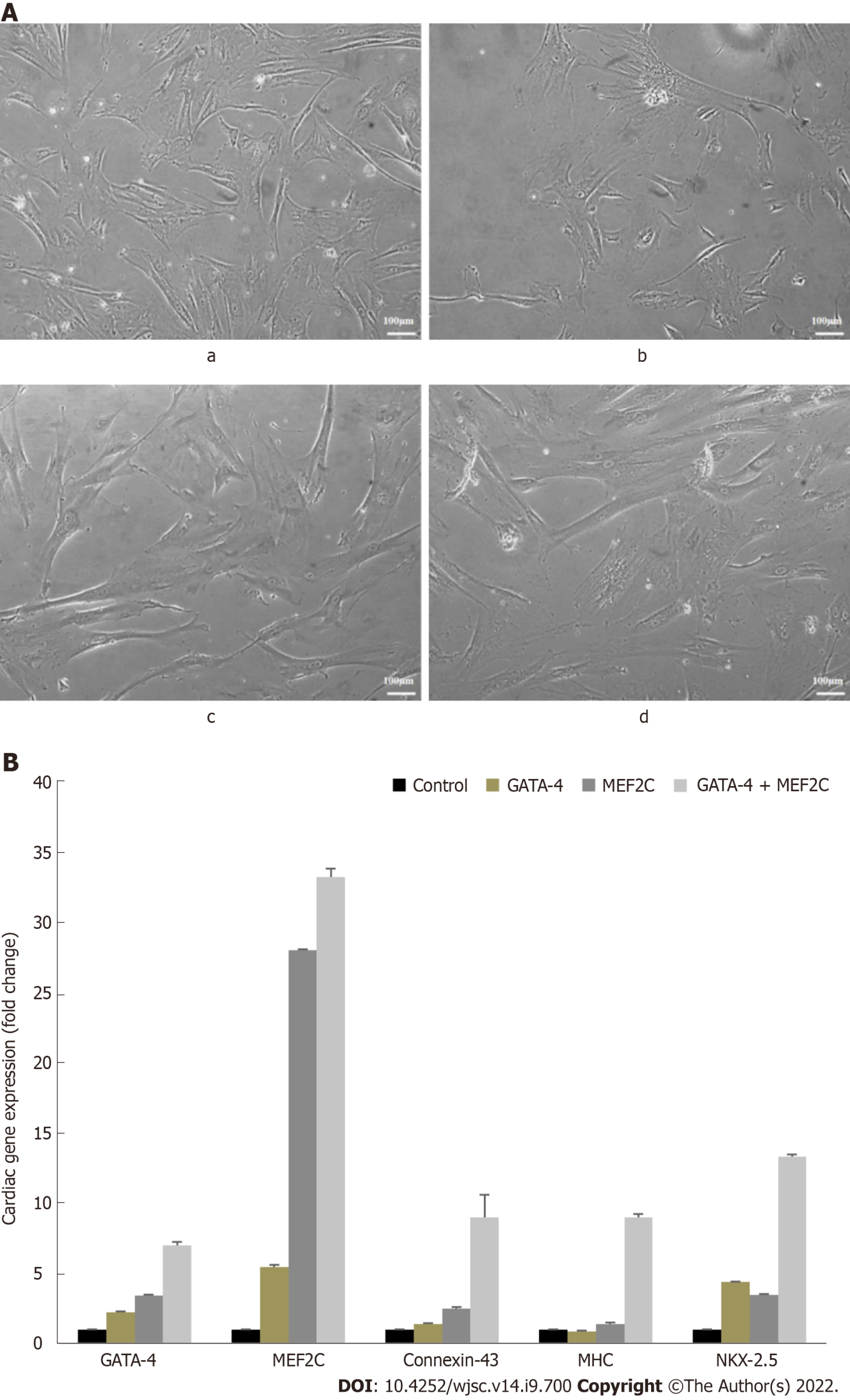
Figure 4 Morphological changes and cardiac-specific gene expression in transfected human umbilical cord mesenchymal stem cells.
A: Images showing human umbilical cord mesenchymal stem cells trasnfected with (b) GATA binding protein 4 (GATA-4), (c) myocyte enhancer factor 2C (MEF2C), and (d) GATA-4 + MEF2C, and (a) the corresponding untreated control. All images were captured at day 14 under a phase contrast microscope (scale bar: 100 μm); B: Bar diagrams showing fold change analysis of cardiac gene expression by semiquantitative real-time polymerase chain reaction (RT-PCR) in the transfected cells in comparison to the control cells after 14 d of culture. Results are expressed as the mean ± SE (n = 3). Differences between groups are considered statistically significant where aP < 0.05, bP < 0.01, and cP < 0.001. GATA-4: GATA binding protein 4; MEF2C: Myocyte enhancer factor 2C; MHC: Myosin heavy chain; NKX2.5: NK2 homeobox 5.
- Citation: Razzaq SS, Khan I, Naeem N, Salim A, Begum S, Haneef K. Overexpression of GATA binding protein 4 and myocyte enhancer factor 2C induces differentiation of mesenchymal stem cells into cardiac-like cells. World J Stem Cells 2022; 14(9): 700-713
- URL: https://www.wjgnet.com/1948-0210/full/v14/i9/700.htm
- DOI: https://dx.doi.org/10.4252/wjsc.v14.i9.700


