
















©The Author(s) 2021.
World J Stem Cells. Feb 26, 2021; 13(2): 177-192
Published online Feb 26, 2021. doi: 10.4252/wjSC.v13.i2.177
Published online Feb 26, 2021. doi: 10.4252/wjSC.v13.i2.177
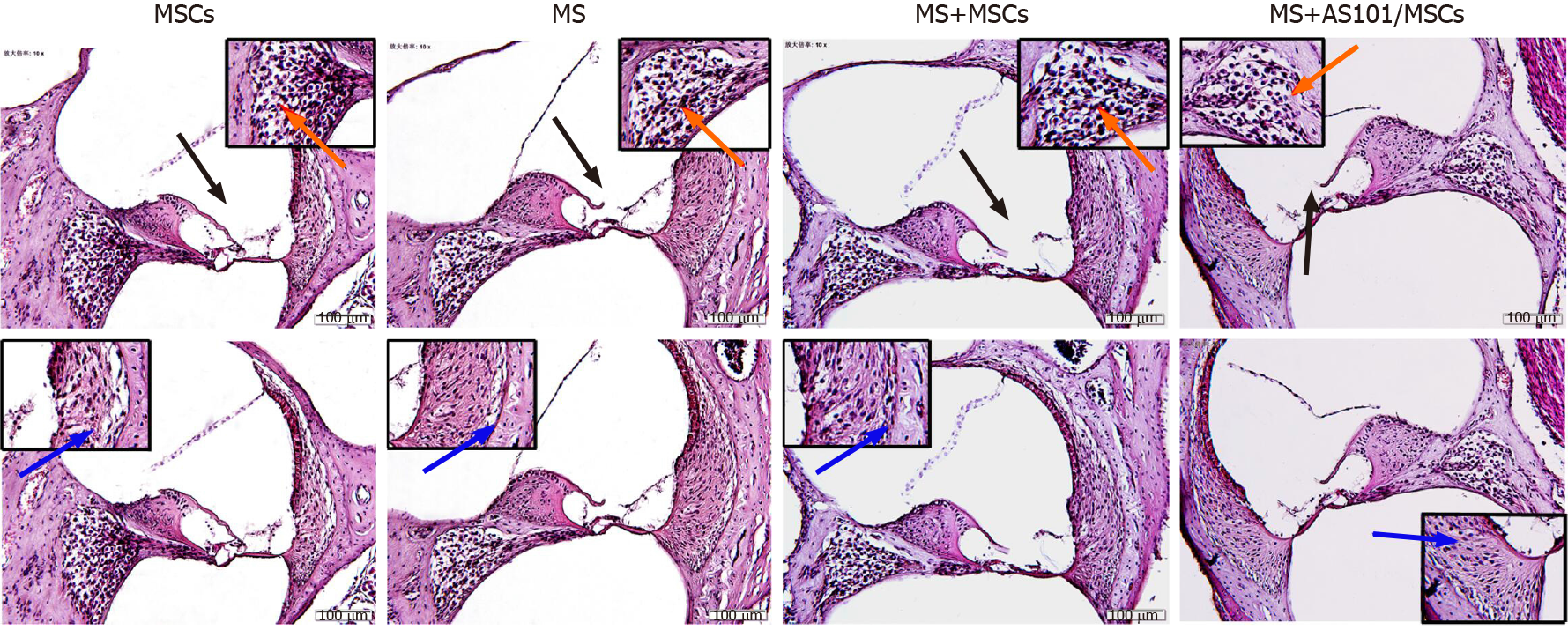
Figure 6 Representative histology images (hematoxylin and eosin staining) of the cochleae in the four groups.
Black arrows indicate the organ of Corti, blue arrows indicate vascular striate cells, and orange arrows indicate spiral ganglion cells. The dotted boxes are featured in the magnified inserts. Images of the four groups are shown in high-power fields (bar, 100 μm). MS: Motion sickness; MSCs: Mesenchymal stem cells.
- Citation: Zhu HS, Li D, Li C, Huang JX, Chen SS, Li LB, Shi Q, Ju XL. Prior transfusion of umbilical cord mesenchymal stem cells can effectively alleviate symptoms of motion sickness in mice through interleukin 10 secretion. World J Stem Cells 2021; 13(2): 177-192
- URL: https://www.wjgnet.com/1948-0210/full/v13/i2/177.htm
- DOI: https://dx.doi.org/10.4252/wjSC.v13.i2.177