Copyright
©The Author(s) 2020.
World J Stem Cells. Aug 26, 2020; 12(8): 706-720
Published online Aug 26, 2020. doi: 10.4252/wjsc.v12.i8.706
Published online Aug 26, 2020. doi: 10.4252/wjsc.v12.i8.706
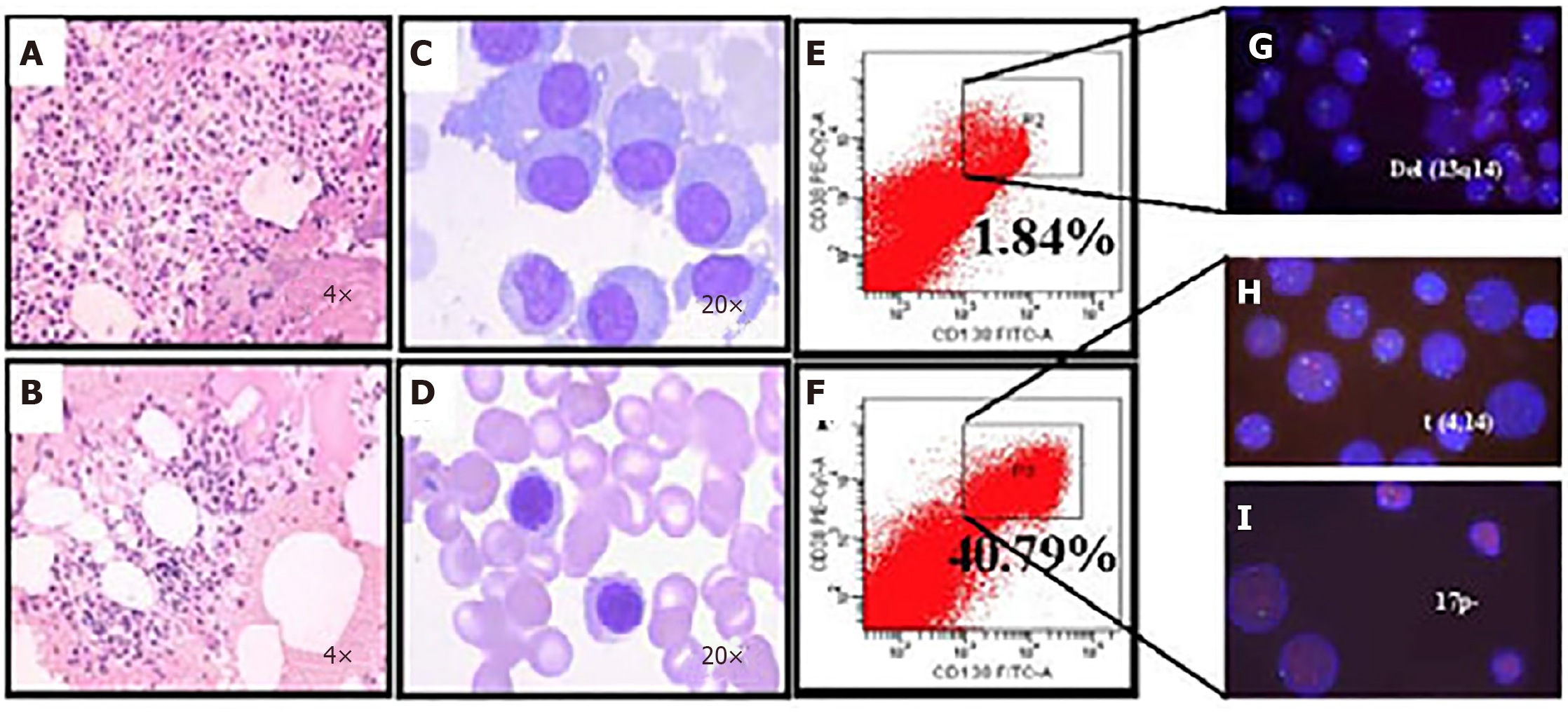
Figure 5 Microfluidic risk-stratification improves clinical outcomes of multiple myeloma (Patient 2).
A: Bone marrow at diagnosis. Active granulocyte hyperplasia; B: Partial remission was achieved with revised risk-stratification; C: At diagnosis, bone marrow plasma cell (PC) abnormalities included clustered and scattered distribution of primitive and immature PCs, with large cell body, fine chromatin, visible nucleolus, and abundant cytoplasm; D: After treatment for high-risk multiple myeloma, PCs were rare and had normal morphology; no typical abnormal PCs were observed; E: At diagnosis without microfluidic enrichment, PCs (CD38+/CD138+) were only 1.84 %; F: After microfluidic enrichment, PCs (CD38+/CD138+) increased to 40.79%; G: Without microfluidic enrichment, FISH showed IgH rearrangement and del(13q14), leading to classification as intermediate risk (Red: D13S319; Green: P53); H and I: After microfluidic enrichment, in addition to del(13q14), FISH showed t (4,14) fusion (yellow dots) and 17p- (Red: D13S319; Green: P53), patient was reclassified and treated as high-risk, which led to efficacious treatment (Refer to[17] for details).
- Citation: Lee LX, Li SC. Hunting down the dominating subclone of cancer stem cells as a potential new therapeutic target in multiple myeloma: An artificial intelligence perspective. World J Stem Cells 2020; 12(8): 706-720
- URL: https://www.wjgnet.com/1948-0210/full/v12/i8/706.htm
- DOI: https://dx.doi.org/10.4252/wjsc.v12.i8.706


