Copyright
©The Author(s) 2023.
World J Stem Cells. Dec 26, 2023; 15(12): 1093-1103
Published online Dec 26, 2023. doi: 10.4252/wjsc.v15.i12.1093
Published online Dec 26, 2023. doi: 10.4252/wjsc.v15.i12.1093
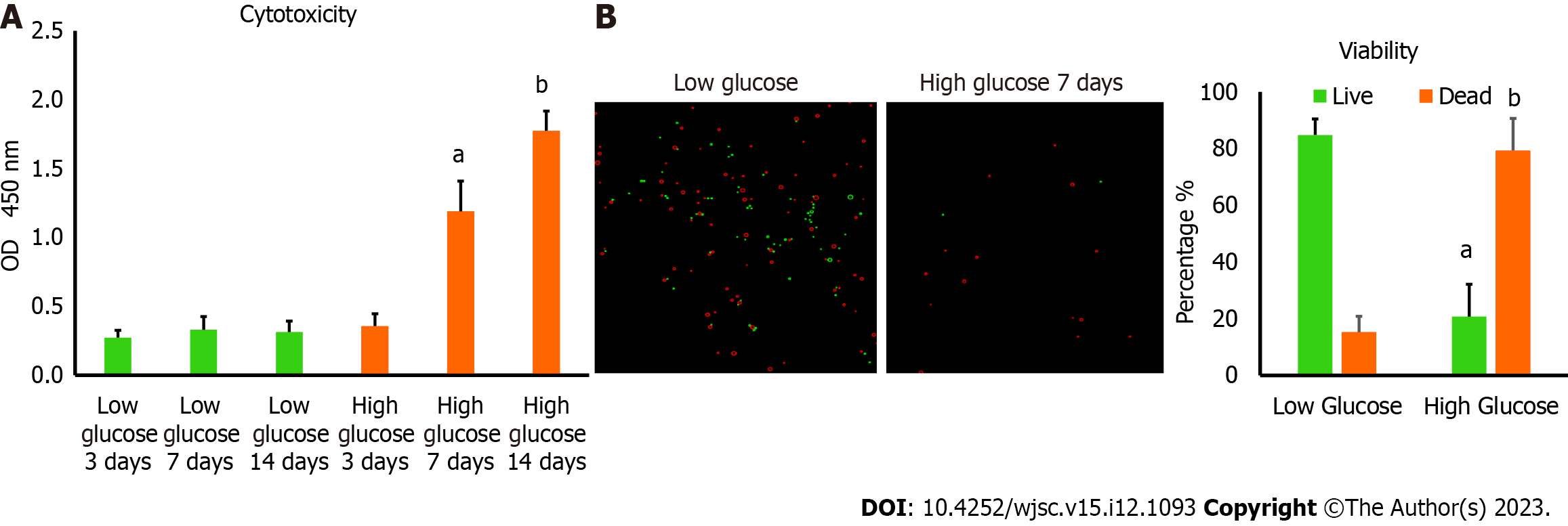
Figure 1 Culturing human adipose tissue-derived mesenchymal stem cells in high glucose reduced their viability.
A: Lactate dehydrogenase cytotoxicity assay revealed higher level of cytotoxicity in human adipose tissue-derived mesenchymal stem cells (hAD-MSCs) cultured in high glucose for 7 and 14 d compared to cells cultured in low glucose. n = 5, aP < 0.01 compared to low glucose 7 d, bP < 0.01 compared to low glucose 14 d; B: The percentage of cells viability detected by 0.4% Trypan blue showed significant reduction in the number of viable hAD-MSCs after being cultured in high glucose for 7 d. Fluorescent images of live and dead cells were obtained using Corning® CytoSmart Cell Counter. n = 5. n = 5, aP < 0.01 compared to live cells in low glucose 7 d, bP < 0.01 compared to dead cells in low glucose 7 d.
Figure 2 High glucose decreased the mitochondrial membrane potential level of human adipose tissue-derived mesenchymal stem cells.
The tetramethylrhodamine ethyl ester (TMRE) fluorescence intensity values of human adipose tissue-derived mesenchymal stem cells (hAD-MSCs) cultured in high glucose for 7 d were substantially declined compared to fluorescent intensity values of cells cultured in low glucose. Live fluorescent images showed a weak signal of TMRE stain in hAD-MSCs after being placed in high glucose for 7 d compared to strong TMRE signal in low glucose cultured cells. n = 4, aP < 0.05 compared to low glucose control. Fluroscnet images were captured at Texas Red filter using Cytation 5 (BioTek, United States). TMRE: Tetramethylrhodamine ethyl ester; RFU: Relative fluorescence units.
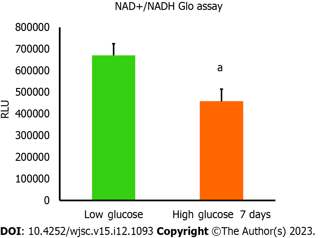
Figure 3 Nicotinamide adenine dinucleotide and NADH amounts were declined in high glucose cultured human adipose tissue-derived mesenchymal stem cells.
The amount of nicotinamide adenine dinucleotide (NAD+) produced from NADH was measured using NAD+/NADH Glo assay. Luminescence intensity was significantly less in human adipose tissue-derived mesenchymal stem cells cultured in high glucose for 7 d compared to cells cultured in low glucose. n = 4, aP < 0.01 compared to low glucose control. Luminescence values were measured using Cytation 5 (BioTek, United States). RFU: Relative fluorescence units.
Figure 4 The level of apoptosis was higher in human adipose tissue-derived mesenchymal stem cells cultured in high glucose.
RealTime-Glo™ Annexin V Apoptosis live assay was used. The level of apoptosis was remarkably increased in human adipose tissue-derived mesenchymal stem cells cultured in high glucose for 7 d. Green fluorescence intensity is corresponding to the number of cells undergoing apoptosis. Fluorescent images were captured at GFP filter using Cytation 5 (BioTek, United States). n = 3, aP < 0.05 compared to low glucose control.
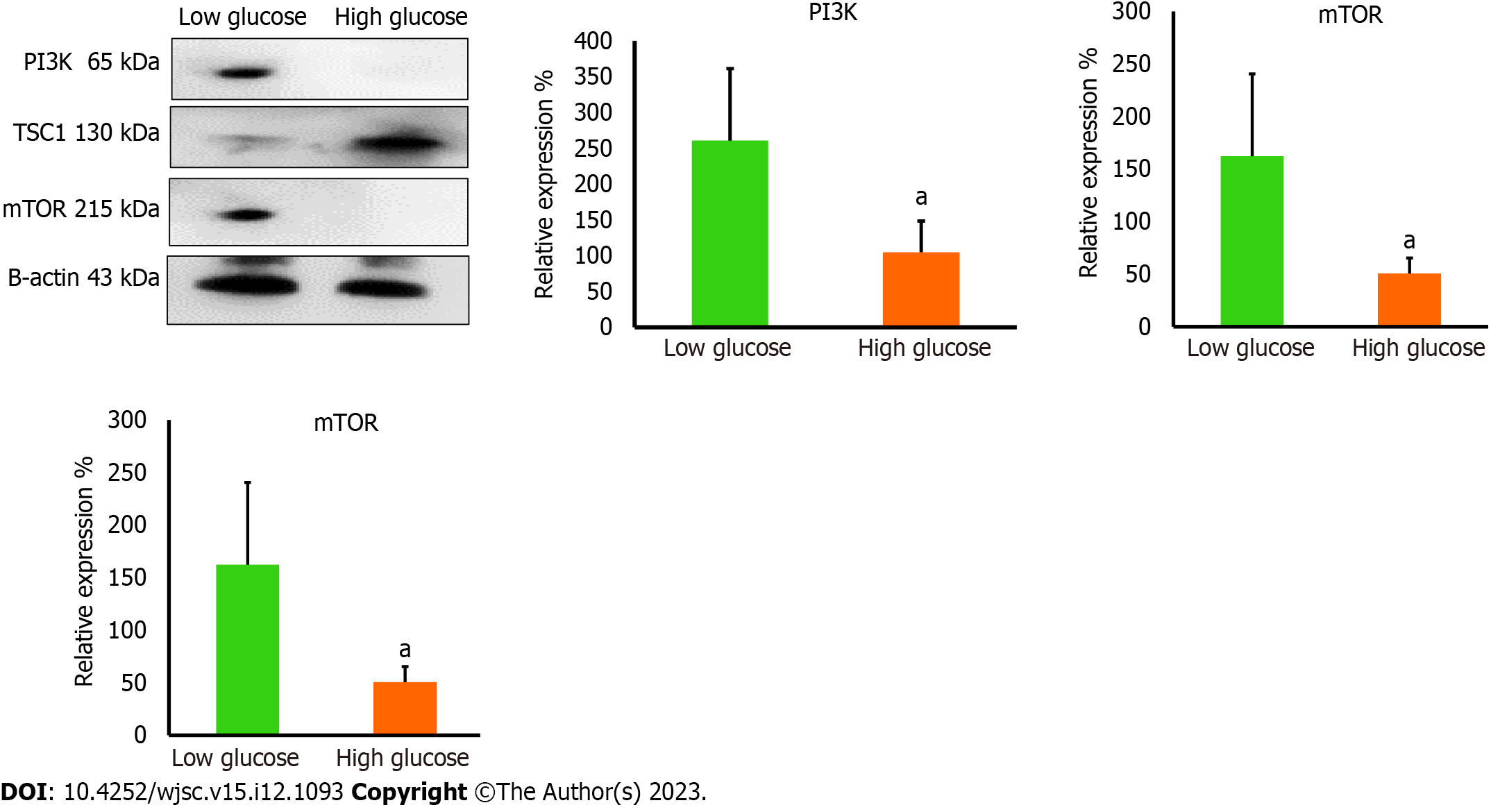
Figure 5 The protein levels of mitochondrial regulators were reduced in high glucose cultured human adipose tissue-derived mesenchymal stem cells.
Western blot protein analysis showed significant reduction in the protein levels of phosphatidylinositol 3-kinase and mammalian target of rapamycin (mTOR) in high glucose cultured cells, while the protein level of mTOR inhibitory protein; TSC1, was elevated in high glucose cultured cells. These proteins are important to maintain proper mitochondrial dynamics. n = 3, aP < 0.05 compared to low glucose control. PI3K: Phosphatidylinositol 3-kinase; mTOR: Mammalian target of rapamycin.
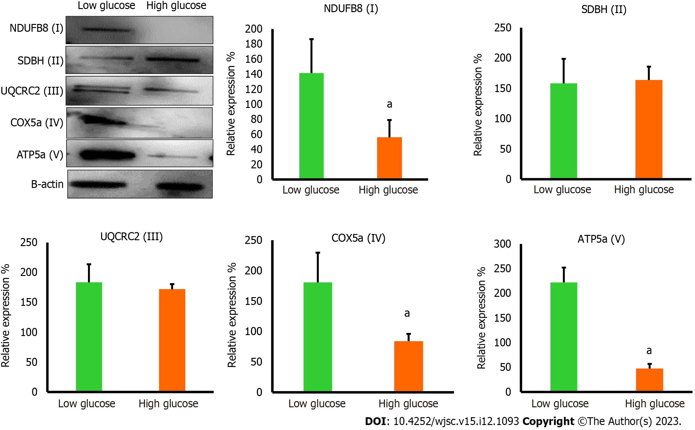
Figure 6 The protein levels of mitochondrial complexes were reduced in high glucose cultured human adipose tissue-derived mesenchymal stem cells.
Western blot protein analysis showed significant reduction in the protein levels of NDUF6B (complex I), COX5a (complex IV), and ATP5a (complex V) in high glucose cultured human adipose tissue-derived mesenchymal stem cells comparing to low glucose cultured cells, while the protein levels of SDBH (complex II) and UQCRC2 (complex III) were not significantly changed in both culturing conditions. n = 3, aP < 0.05 compared to low glucose control.
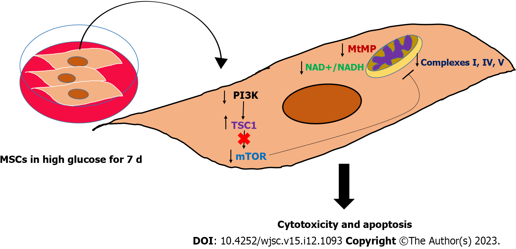
Figure 7 Graphical summary of the study findings.
High glucose impaired the mitochondrial function in mesenchymal stem cells (MSCs) by perturbing many mitochondrial regulatory dynamics and factors, particularly mitochondrial membrane potential, nicotinamide adenine dinucleotide (NAD+)/NADH pool, and mammalian target of rapamycin protein. This mitochondrial dysfunction is associated with triggering apoptosis in MSCs, which mechanistically explained the poor survival of MSCs in hyperglycemia. MSCs: Mesenchymal stem cells; PI3K: Phosphatidylinositol 3-kinase; mTOR: Mammalian target of rapamycin; NAD+: Nicotinamide adenine dinucleotide.
- Citation: Abu-El-Rub E, Almahasneh F, Khasawneh RR, Alzu'bi A, Ghorab D, Almazari R, Magableh H, Sanajleh A, Shlool H, Mazari M, Bader NS, Al-Momani J. Human mesenchymal stem cells exhibit altered mitochondrial dynamics and poor survival in high glucose microenvironment. World J Stem Cells 2023; 15(12): 1093-1103
- URL: https://www.wjgnet.com/1948-0210/full/v15/i12/1093.htm
- DOI: https://dx.doi.org/10.4252/wjsc.v15.i12.1093


