Copyright
©The Author(s) 2019.
World J Stem Cells. Jan 26, 2019; 11(1): 1-12
Published online Jan 26, 2019. doi: 10.4252/wjsc.v11.i1.1
Published online Jan 26, 2019. doi: 10.4252/wjsc.v11.i1.1
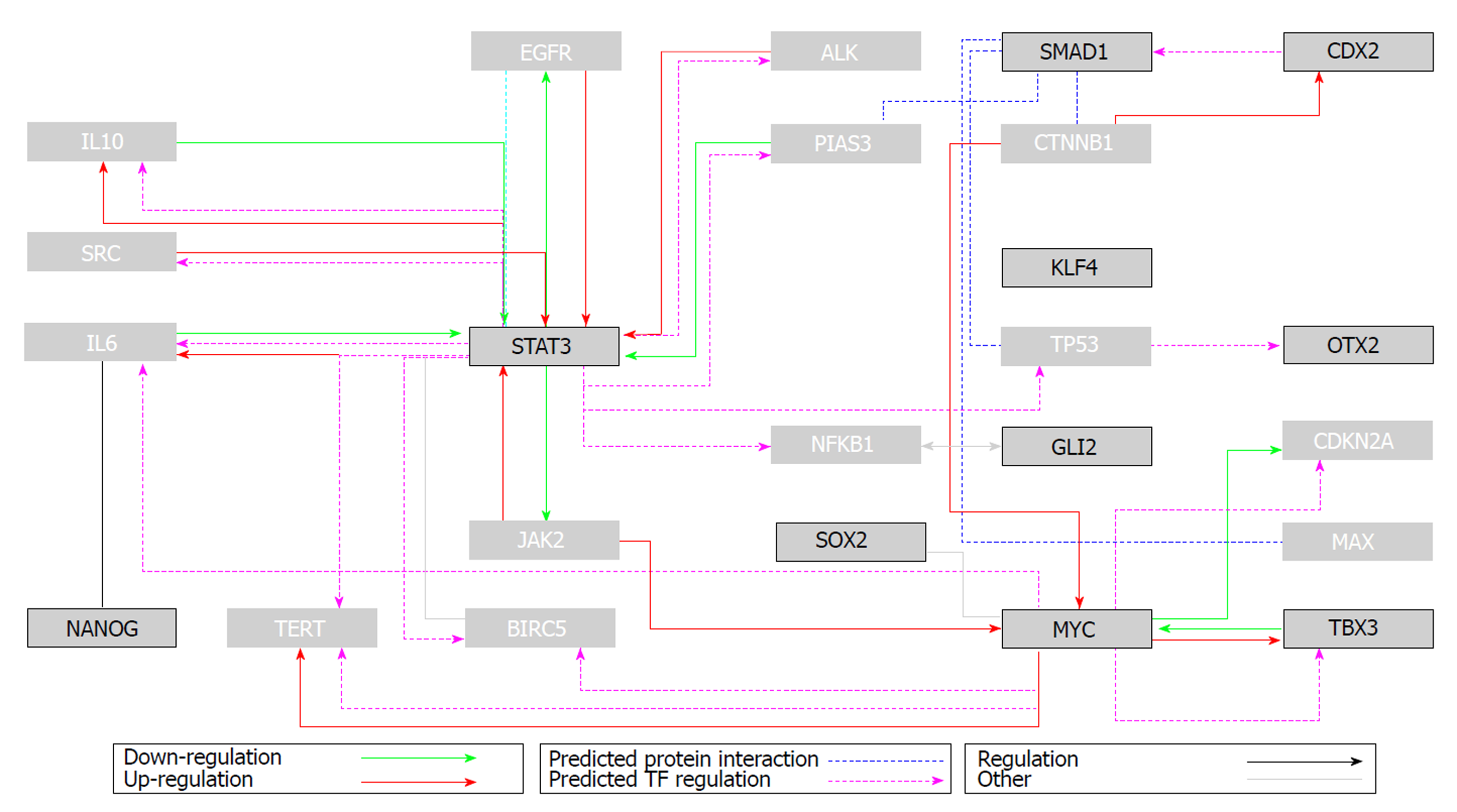
Figure 1 Flux diagram of the top 10 ranked genes related to pluripotency (interaction data obtained from GNCPro, SABiosciences).
Interactions: downregulation (green arrow), upregulation (red arrow), predicted transcription factor regulation (magenta arrow), predicted protein interaction (blue line), regulation (black arrow), other types of regulation (grey line). See the electronic version for colour figures. Boxes outlined in black represent the target genes, and light grey boxes their immediate neighbours. Adapted from Mashayekhi et al[11].
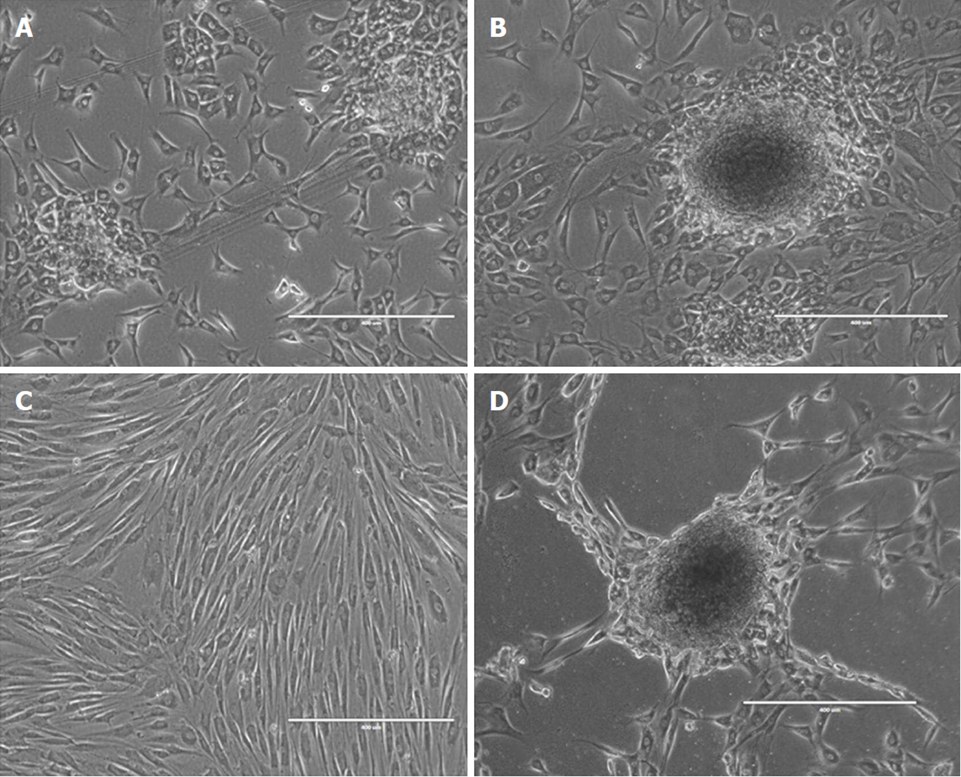
Figure 2 The possibility that severe stress, cell loss or even ageing can induce human adult pluripotent cells both in vitro and in vivo cannot be ruled out and should warrant further investigation.
A: A normal human dermal fibroblast (NHDF) cell line after 48 h of intentional CO2 absence in the incubator. The cells modified their morphologic characteristics and adopted the culture appearance of pluripotent cells; B: Modified NHDF cells 28 d after stress; C: Non-exposed NHDF cells; D: Modified NHDF cells 73 d after stress.
- Citation: Labusca L, Mashayekhi K. Human adult pluripotency: Facts and questions. World J Stem Cells 2019; 11(1): 1-12
- URL: https://www.wjgnet.com/1948-0210/full/v11/i1/1.htm
- DOI: https://dx.doi.org/10.4252/wjsc.v11.i1.1


