Copyright
©The Author(s) 2003.
World J Gastroenterol. Aug 15, 2003; 9(8): 1799-1803
Published online Aug 15, 2003. doi: 10.3748/wjg.v9.i8.1799
Published online Aug 15, 2003. doi: 10.3748/wjg.v9.i8.1799
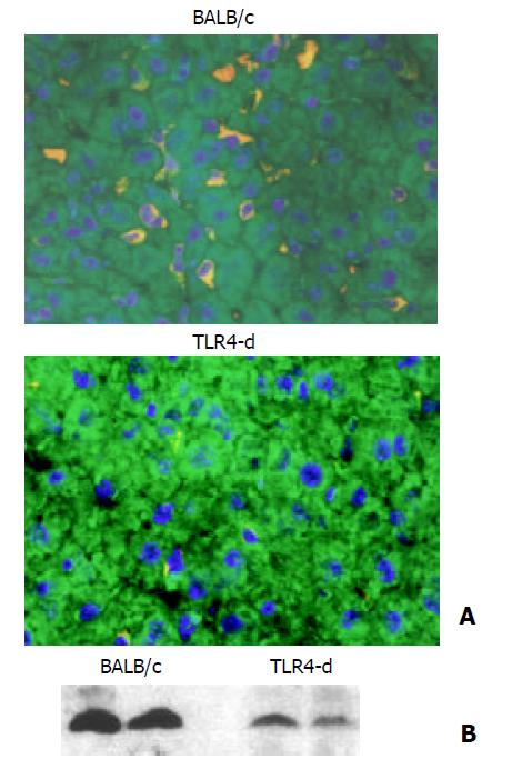
Figure 3 Effect of TLR4 mutation on liver HO-1 expression.
A: Immunofluorescent detection of HO-1 in liver. After three doses of LPS treatment, liver HO-1 expression was visualized by immunofluorescent staining with a specific rabbit polyclonal antibody against HO-1, followed by indocarbocyanine (Cy3)-conjugated anti-rabbit IgG (red). The cell surface was counter-stained with fluorescein-conjugated wheat germ agglutinin (green), and the nucleus was counterstained with bis-benzimide (blue). Liver HO-1 expression was abrogated in TLR4 mutated mice following LPS, but neither was influenced in BALB/c (control) mice (magnification ×400). B: Immunoblotting detection of HO-1 in liver. BALB/c and TLR4 mutated mice were treated with three doses of LPS. Liver tissue was homogenized, and immunoblotting analysis was performed. HO-1 protein was detected by immunoblotting with polyclonal rabbit antibody against HO-1. Data were repre-sentative of at least 2 experiments.
- Citation: Song Y, Shi Y, Ao LH, Harken AH, Meng XZ. TLR4 mediates LPS-induced HO-1 expression in mouse liver: Role of TNF-α and IL-1β. World J Gastroenterol 2003; 9(8): 1799-1803
- URL: https://www.wjgnet.com/1007-9327/full/v9/i8/1799.htm
- DOI: https://dx.doi.org/10.3748/wjg.v9.i8.1799


