Copyright
©The Author(s) 2003.
World J Gastroenterol. Aug 15, 2003; 9(8): 1667-1674
Published online Aug 15, 2003. doi: 10.3748/wjg.v9.i8.1667
Published online Aug 15, 2003. doi: 10.3748/wjg.v9.i8.1667
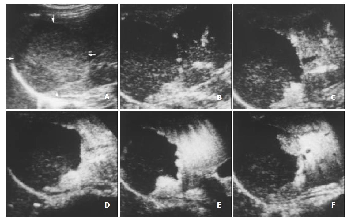
Figure 4 A 45-year-old female with hemangioma.
A. Intercostal section of precontrast conventional sonography exhibits a hypoechogenic lesion (arrows) of 6.5 cm in diameter. B. Contrast-enhanced C-cube gray scale sonography at 23 s (B) after injection of Levovist shows that no microbubble signal appears in the lesion while enhancement in the liver parenchyma begins. C-F. C-cube gray scale sonography demonstrates gradual peripheral enhancement at 32 s (C), 47 s (D), 113 s (E) and 370 s (F) with a long enhancement duration.
- Citation: Wang WP, Ding H, Qi Q, Mao F, Xu ZZ, Kudo M. Characterization of focal hepatic lesions with contrast-enhanced C-cube gray scale ultrasonography. World J Gastroenterol 2003; 9(8): 1667-1674
- URL: https://www.wjgnet.com/1007-9327/full/v9/i8/1667.htm
- DOI: https://dx.doi.org/10.3748/wjg.v9.i8.1667


