Copyright
©The Author(s) 2003.
World J Gastroenterol. Aug 15, 2003; 9(8): 1667-1674
Published online Aug 15, 2003. doi: 10.3748/wjg.v9.i8.1667
Published online Aug 15, 2003. doi: 10.3748/wjg.v9.i8.1667
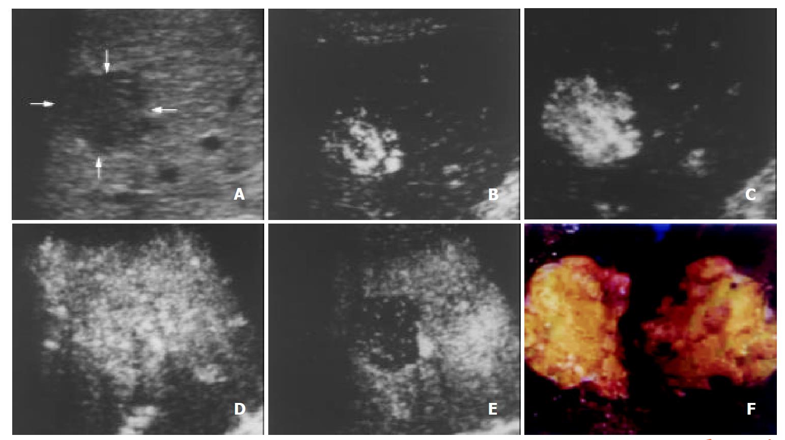
Figure 1 A 47-year-old man with hepatocellular carcinoma.
A. Intercostal precontrast conventional sonography exhibits a hypoechogenic lesion (arrows) of 2.7 cm in diameter. B-E. Contrast-enhanced C-cube gray scale sonography at 23 s (B), 28 s (C) and 43 s (D) after injection of Levovist shows that intranodular signals enhance gradually and earlier than the liver parenchyma, and enhancement decreases rapidly in the portal venous phase (110 s, E). This suggests hypervascularity of hepatocellular carcinoma with an early enhancement and early wash-out of contrast. F. Gross specimen of the resected tumor exhibits a gray fish-like profile and suggests the typical appearance of hepatocellular carcinoma.
- Citation: Wang WP, Ding H, Qi Q, Mao F, Xu ZZ, Kudo M. Characterization of focal hepatic lesions with contrast-enhanced C-cube gray scale ultrasonography. World J Gastroenterol 2003; 9(8): 1667-1674
- URL: https://www.wjgnet.com/1007-9327/full/v9/i8/1667.htm
- DOI: https://dx.doi.org/10.3748/wjg.v9.i8.1667


