Copyright
©The Author(s) 2003.
World J Gastroenterol. Jul 15, 2003; 9(7): 1398-1403
Published online Jul 15, 2003. doi: 10.3748/wjg.v9.i7.1398
Published online Jul 15, 2003. doi: 10.3748/wjg.v9.i7.1398
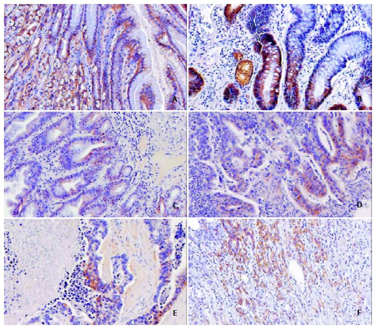
Figure 2 Immunohistochemical staining of CA IX in normal gastric mucosa (A), metaplasia (B), adenoma with moderate dysplasia (C), adenocarcinoma grade II (D), adenocarcinoma grade III (E), and diffuse carcinoma (F).
In the normal mucosa, CA IX was expressed in all histological layers covering the luminal surface (LS), proliferative zone (PZ), and gastric glands (G). It was notable that CA IX was confined to deep crypts in metaplastic epithelium. The adenoma sample and all malignant lesions shown in this figure were positive for CA IX. (Original magnifications × 200).
- Citation: Leppilampi M, Saarnio J, Karttunen TJ, Kivelä J, Pastoreková S, Pastorek J, Waheed A, Sly WS, Parkkila S. Carbonic anhydrase isozymes IX and XII in gastric tumors. World J Gastroenterol 2003; 9(7): 1398-1403
- URL: https://www.wjgnet.com/1007-9327/full/v9/i7/1398.htm
- DOI: https://dx.doi.org/10.3748/wjg.v9.i7.1398


