Copyright
©The Author(s) 2003.
World J Gastroenterol. Apr 15, 2003; 9(4): 813-817
Published online Apr 15, 2003. doi: 10.3748/wjg.v9.i4.813
Published online Apr 15, 2003. doi: 10.3748/wjg.v9.i4.813
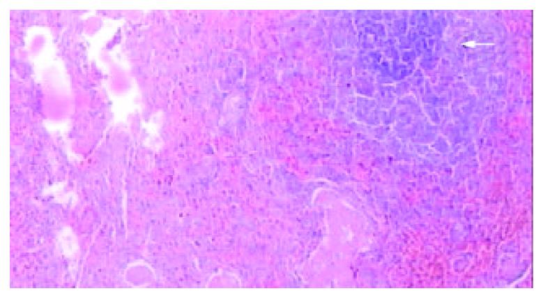
Figure 9 Microscopic appearance of “plenic carnification”of the normal viable splenic tissue distant form ablative lesion at the end of 9th week after RFA, the tissue structure consolidated, larger vessels occluded, extensive fibrous protein deposited, and the congestive splenic sinusoid disappeared; however, the struc-ture of splenic lymphoid nodule was intact (arrow) (HE.
× 40).
- Citation: Liu QD, Ma KS, He ZP, Ding J, Huang XQ, Dong JH. Experimental study on the feasibility and safety of radiofrequency ablation for secondary splenomagely and hypersplenism. World J Gastroenterol 2003; 9(4): 813-817
- URL: https://www.wjgnet.com/1007-9327/full/v9/i4/813.htm
- DOI: https://dx.doi.org/10.3748/wjg.v9.i4.813


