Copyright
©The Author(s) 2003.
World J Gastroenterol. Apr 15, 2003; 9(4): 813-817
Published online Apr 15, 2003. doi: 10.3748/wjg.v9.i4.813
Published online Apr 15, 2003. doi: 10.3748/wjg.v9.i4.813
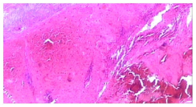
Figure 6 Light microscopic appearance of the coagulative ne-crosis at the end of 2nd week after RFA, the intrasplenic hemor-rhage at the probe insertion site could see (arrow), the splenic capsule thickened (HE.
× 40).
- Citation: Liu QD, Ma KS, He ZP, Ding J, Huang XQ, Dong JH. Experimental study on the feasibility and safety of radiofrequency ablation for secondary splenomagely and hypersplenism. World J Gastroenterol 2003; 9(4): 813-817
- URL: https://www.wjgnet.com/1007-9327/full/v9/i4/813.htm
- DOI: https://dx.doi.org/10.3748/wjg.v9.i4.813


