Copyright
©The Author(s) 2003.
World J Gastroenterol. Dec 15, 2003; 9(12): 2751-2758
Published online Dec 15, 2003. doi: 10.3748/wjg.v9.i12.2751
Published online Dec 15, 2003. doi: 10.3748/wjg.v9.i12.2751
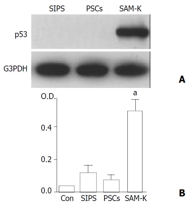
Figure 3 p53 expression and telomerase activity were nega-tive in SIPS cells.
(A) Total cell lysates (approximately 100 μg) were prepared from SIPS, primary PSCs (passage 3), and SAM-K cells. The level of p53 was determined by Western blotting. The level of G3PDH was also determined as a loading control. (B) Telomerase activity was measured utilizing the TRAP by enzyme-linked immunosorbent assay. The telomerase activity was determined by differences in absorbance at O.D.450-O.D. 690. Negative control was prepared by incubating the total cell extracts with ribonuclease A. The mean of the absorbance in negative control was subtracted from those of the samples, and the samples were regarded as telomerase positive (“a”) if the difference in absorbance was higher than 0.2. Data are shown as mean ± SD (n = 6).
- Citation: Masamune A, Satoh M, Kikuta K, Suzuki N, Shimosegawa T. Establishment and characterization of a rat pancreatic stellate cell line by spontaneous immortalization. World J Gastroenterol 2003; 9(12): 2751-2758
- URL: https://www.wjgnet.com/1007-9327/full/v9/i12/2751.htm
- DOI: https://dx.doi.org/10.3748/wjg.v9.i12.2751


