Copyright
©The Author(s) 2003.
World J Gastroenterol. Oct 15, 2003; 9(10): 2198-2201
Published online Oct 15, 2003. doi: 10.3748/wjg.v9.i10.2198
Published online Oct 15, 2003. doi: 10.3748/wjg.v9.i10.2198
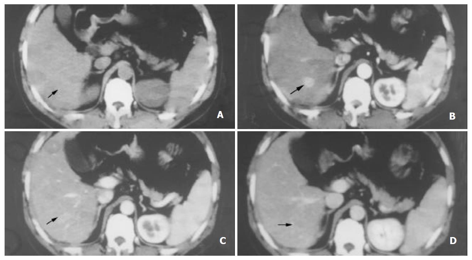
Figure 3 SHCC of 1.
5 cm in diameter, A: Hypoattenuating lesion showed in plance image. B: CT scan obtained at the same level as in the early arterial phase, Tumor intensely enhanced. C: In the late arterial phase tumor becomes isodense (arrow). D: In the portal venous phase, the tumor turned to hypodense.
- Citation: Zhao H, Zhou KR, Yan FH. Role of multiphase scans by multirow-detector helical CT in detecting small hepatocellular carcinoma. World J Gastroenterol 2003; 9(10): 2198-2201
- URL: https://www.wjgnet.com/1007-9327/full/v9/i10/2198.htm
- DOI: https://dx.doi.org/10.3748/wjg.v9.i10.2198


