Copyright
©The Author(s) 2002.
World J Gastroenterol. Apr 15, 2002; 8(2): 312-317
Published online Apr 15, 2002. doi: 10.3748/wjg.v8.i2.312
Published online Apr 15, 2002. doi: 10.3748/wjg.v8.i2.312
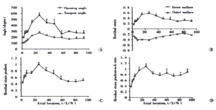
Figure 4 Photographs showing (A) the distributions of the opening angle and tangent rotation angle, (B) residual strains at the mucosal and serosal surfaces, (C) the residual strain gradient and (D) residual strain gradient normalised with respect to the wall thickness (h).
Values are ¯x ± s. A significant variation was found along the small intestine for all variables (P < 0.001).
- Citation: Zhao JB, Sha H, Zhuang FY, Gregersen H. Morphological properties and residual strain along the small intestine in rats. World J Gastroenterol 2002; 8(2): 312-317
- URL: https://www.wjgnet.com/1007-9327/full/v8/i2/312.htm
- DOI: https://dx.doi.org/10.3748/wjg.v8.i2.312


