Copyright
©The Author(s) 2000.
World J Gastroenterol. Aug 15, 2000; 6(4): 540-545
Published online Aug 15, 2000. doi: 10.3748/wjg.v6.i4.540
Published online Aug 15, 2000. doi: 10.3748/wjg.v6.i4.540
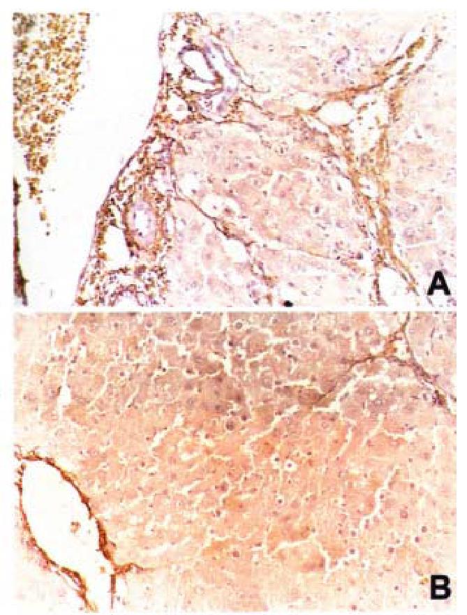
Figure 2 AT1 receptor expression in liver section of model rat (DAB staining, × 200).
AT1 receptor is seen mainly scattered in fibrotic areas and vascular wal l (A), and of los artan treated group (DAB staining, × 200). Note significantly less AT1 expression than that seen in the model group (B).
- Citation: Wei HS, Li DG, Lu HM, Zhan YT, Wang ZR, Huang X, Zhang J, Cheng JL, Xu QF. Effects of AT1 receptor antagonist, losartan, on rat hepatic fibrosis induced by CCl4. World J Gastroenterol 2000; 6(4): 540-545
- URL: https://www.wjgnet.com/1007-9327/full/v6/i4/540.htm
- DOI: https://dx.doi.org/10.3748/wjg.v6.i4.540


