
















©The Author(s) 1998.
World J Gastroenterol. Jun 15, 1998; 4(3): 214-218
Published online Jun 15, 1998. doi: 10.3748/wjg.v4.i3.214
Published online Jun 15, 1998. doi: 10.3748/wjg.v4.i3.214
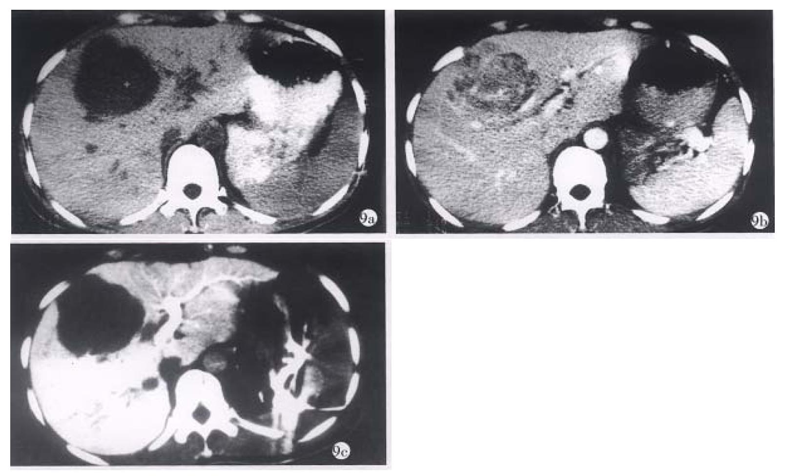
Figure 9 Primary hepatocyte carcinoma of a 30-year-old female patient.
a. plain CT scan; b. enhanced CT; c. CTSP. Though they all could show carcinoma, CTSP not only could show it more clearly than the other two, but also show metastases to the right of the inferior vena cava.
- Citation: Zhang XL, Qiu SJ, Chang RM, Zou CJ. Animal experiments and clinical application of CT during percutaneous splenoportography. World J Gastroenterol 1998; 4(3): 214-218
- URL: https://www.wjgnet.com/1007-9327/full/v4/i3/214.htm
- DOI: https://dx.doi.org/10.3748/wjg.v4.i3.214