
















Copyright: ©Author(s) 2027.
World J Gastroenterol. Jan 7, 2027; 33(1): 122550
Published online Jan 7, 2027. doi: 10.3748/wjg.122550
Published online Jan 7, 2027. doi: 10.3748/wjg.122550
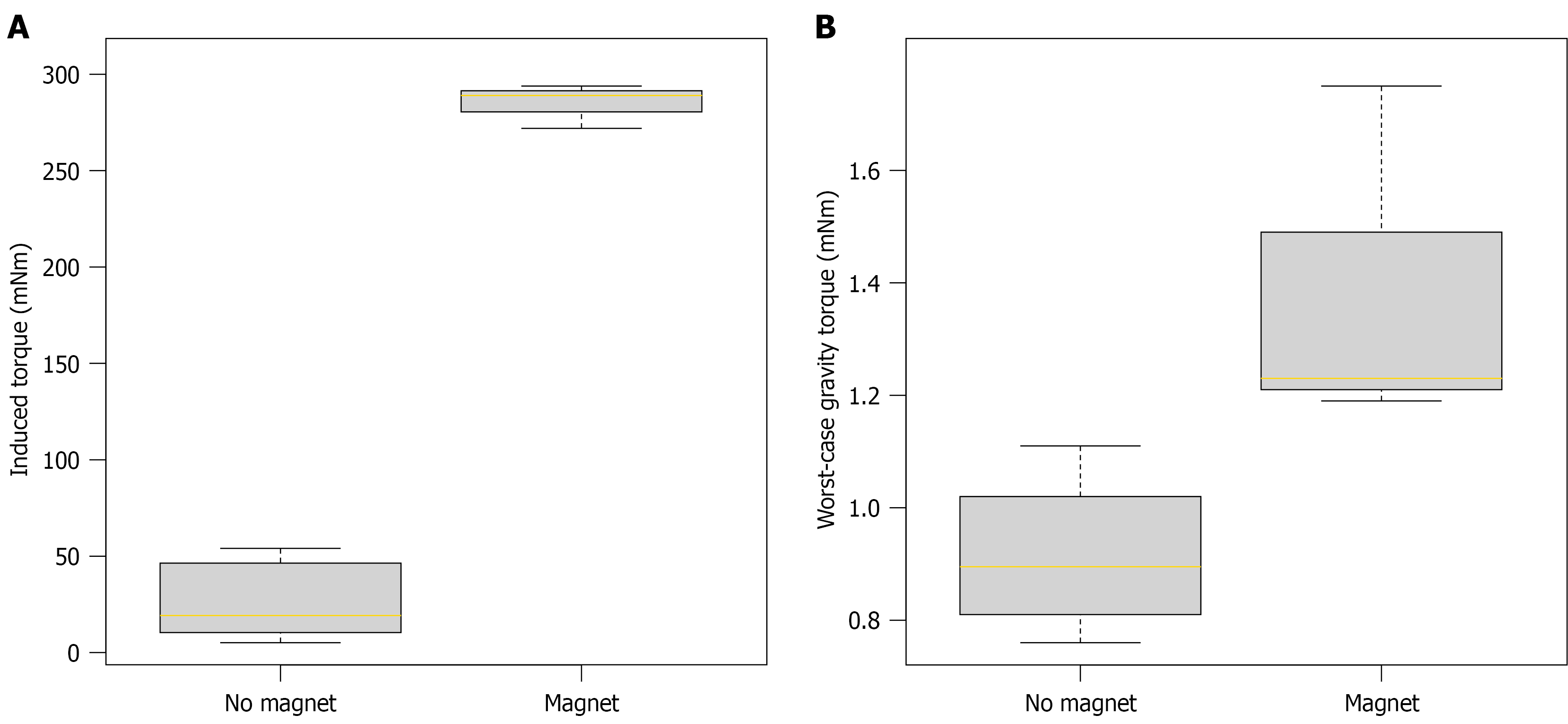
Figure 9 Box plots of magnetically induced torque and gravity-induced torque on video capsule endoscopes.
A: Magnetically induced torque on video capsule endoscopes (VCEs) without an integrated permanent magnet (left; measured) and with an integrated permanent magnet (right; calculated from displacement force); B: Worst-case torque due to gravity on VCEs without (left) and with (right) an integrated permanent magnet. The lengths of the boxes mark the 25th and 75th percentiles, the yellow lines indicate the 50th percentile (median), and the whiskers represent the 1.5-fold interquartile range. Note the different scales of the Y-axis in Figure 9A and B.
- Citation: Beck MJ, Keuchel M, Paschen J, Laser RM, Roling J, Bandorski D, Windhorst AC, Kurniawan N, Baltes P. Magnetic resonance imaging-induced forces on video capsule endoscopes: First systematic in vitro assessment. World J Gastroenterol 2027; 33(1): 122550
- URL: https://www.wjgnet.com/1007-9327/full/v33/i1/122550.htm
- DOI: https://dx.doi.org/10.3748/wjg.122550