
















©The Author(s) 2026.
World J Gastroenterol. Feb 21, 2026; 32(7): 115143
Published online Feb 21, 2026. doi: 10.3748/wjg.v32.i7.115143
Published online Feb 21, 2026. doi: 10.3748/wjg.v32.i7.115143
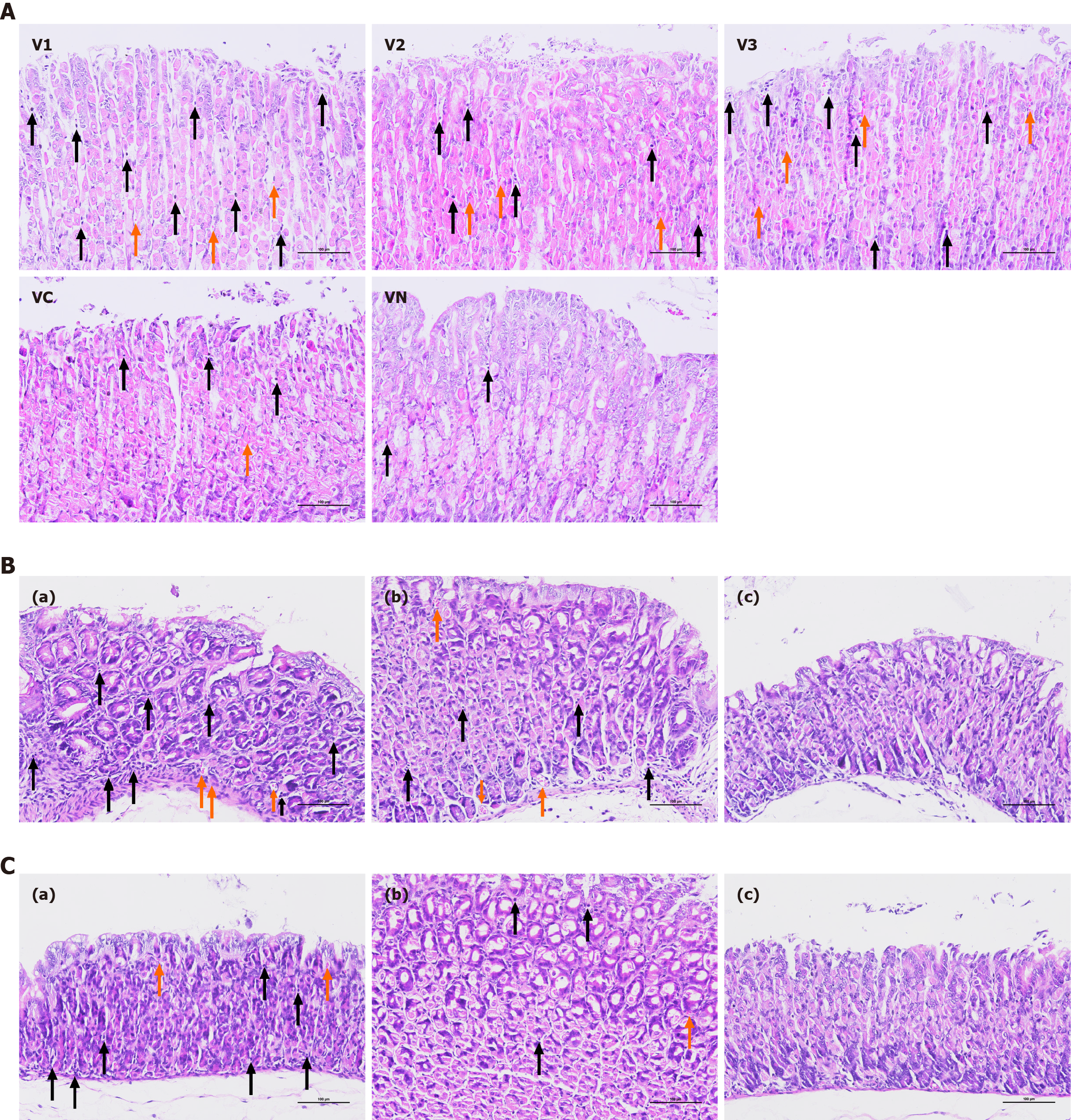
Figure 4 Hematoxylin and eosin staining of the gastric mucosa in Candida vaginal infection model (× 200).
A: Maternal mice; B: Three-week-old offspring mice; C: Six-week-old offspring mice. a: Gastric mucosa from offspring mice in the experimental group; b: Gastric mucosa from offspring in the control group; c: Gastric mucosa from offspring in the normal group. V1: Vagina infected with Candida J115; V2: Vagina infected with Candida H100; V3: Vagina infected with Candida Ca-co-Hp; VC: Vagina infected with Candida Ca10231; VN: Vagina treated with saline solution. The orange and black arrows in the figure indicate the bleeding points and inflammatory cells, respectively.
- Citation: Xu XL, Luo TT, Liu Q, Cui GZ, Wu DY, Wu XJ, Yang TX, Sun JC, Jian HF, Zhuo SW, Chen ZH. Candida-mediated vertical transmission of Helicobacter pylori in C57BL/6J mice. World J Gastroenterol 2026; 32(7): 115143
- URL: https://www.wjgnet.com/1007-9327/full/v32/i7/115143.htm
- DOI: https://dx.doi.org/10.3748/wjg.v32.i7.115143