
















Copyright: ©Author(s) 2026.
World J Gastroenterol. Dec 28, 2026; 32(48): 122778
Published online Dec 28, 2026. doi: 10.3748/wjg.122778
Published online Dec 28, 2026. doi: 10.3748/wjg.122778
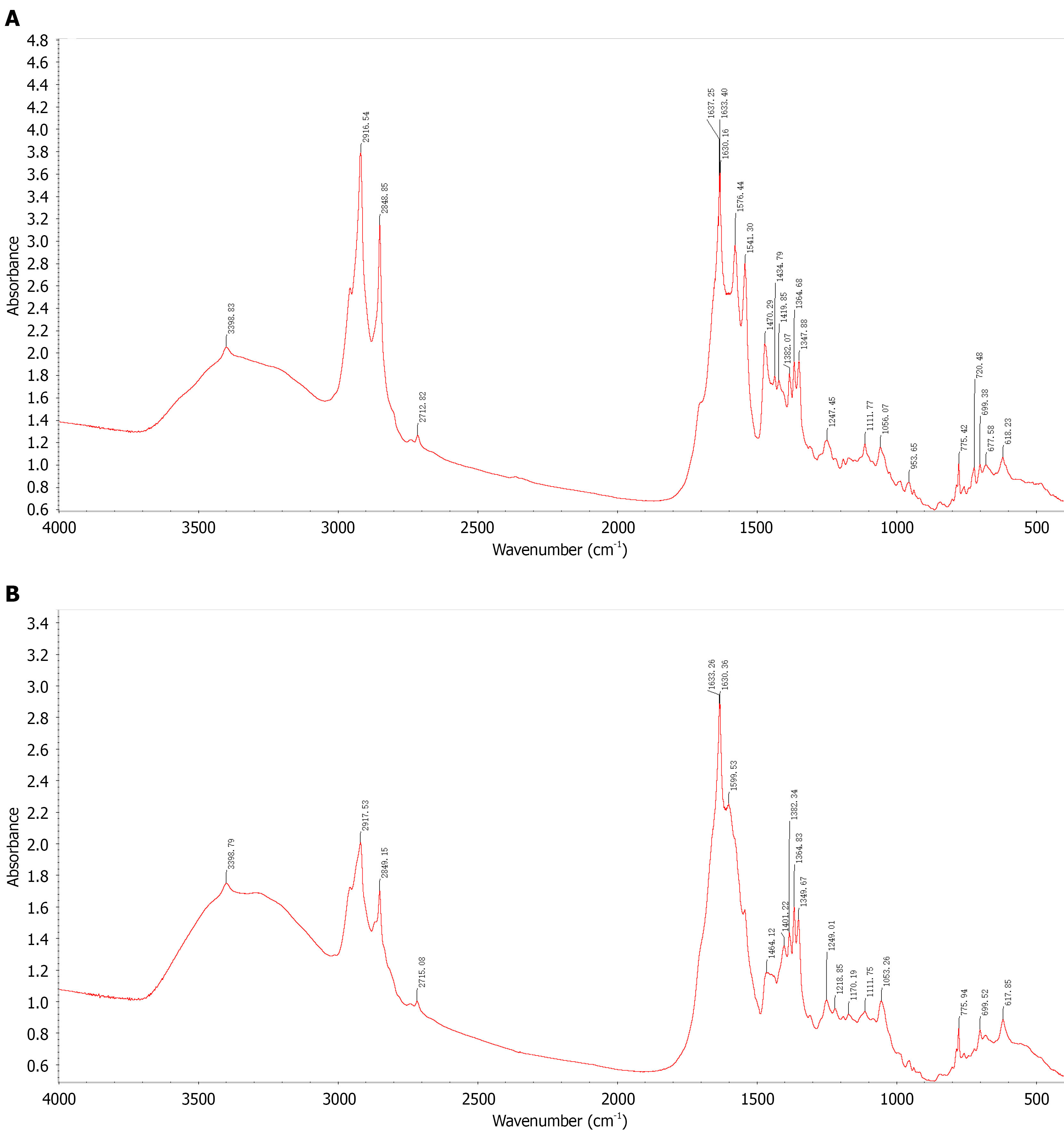
Figure 5 Fourier transform infrared spectroscopy.
A: FT-IR spectrum of clinically confirmed calcium bilirubinate hepatolithiasis specimens. Key absorption bands: Broad peak at 3600-2400 cm-1 (bound water); 3-peak combination at approximately 1637/1633/1630 cm-1, approximately 1576 cm-1, and approximately 1541 cm-1 (characteristic calcium bilirubinate signature); C-H stretching at 2916/2849 cm-1 (cholesterol). This spectrum serves as the reference standard confirming the analytical method; B: FT-IR spectrum of bile sediment from the experimental group (LPS-treated rats). The spectrum closely matches the clinical reference (Figure 5A): Broad bound-water absorption at 3600-2400 cm-1; 3-peak combination near 1633/1630 cm-1, 1599 cm-1, and the 1500-1400 cm-1 region, confirming calcium bilirubinate; C-H stretching at 2917/2849 cm-1, indicating a minor cholesterol component. Taken together, the sediment is predominantly calcium bilirubinate with a small amount of cholesterol. FT-IR: Fourier transform infrared spectroscopy; LPS: Lipopolysaccharide.
- Citation: Shi JL, Muhammad T, He TT, Lin ZY, Zhang TG, Liu SY, Yang XL, Yang Y, Hu L, Ran P, Numan M, Li J. Establishment of a Sprague-Dawley rat model of hepatolithiasis based on common bile duct catheterization. World J Gastroenterol 2026; 32(48): 122778
- URL: https://www.wjgnet.com/1007-9327/full/v32/i48/122778.htm
- DOI: https://dx.doi.org/10.3748/wjg.122778