
















Copyright: ©Author(s) 2026.
World J Gastroenterol. Oct 14, 2026; 32(38): 118814
Published online Oct 14, 2026. doi: 10.3748/wjg.118814
Published online Oct 14, 2026. doi: 10.3748/wjg.118814
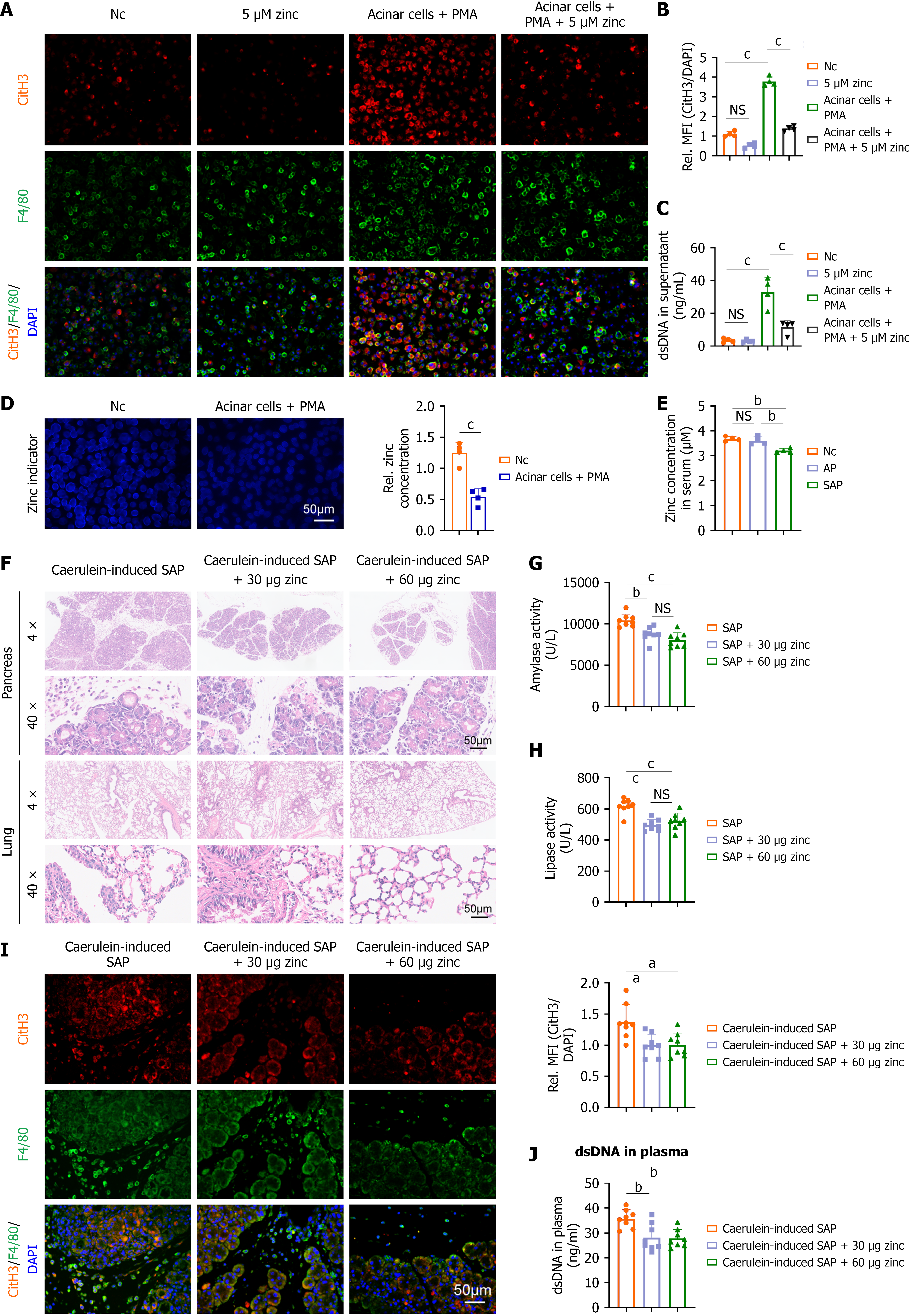
Figure 4 Zinc supplementation can inhibit the formation of macrophage extracellular traps.
A and B: Representative immunofluorescence images of citrullinated histone H3 (CitH3) (red) and F4/80 (green) in macrophages, along with statistical analysis of the relative expression levels of CitH3 (n = 4); C: Double-stranded DNA (dsDNA) levels in the cell supernatant (n = 4); D: Macrophages were cocultured with pancreatic acinar cells treated with caerulein in the presence of phorbol 12-myristate 13-acetate. The intracellular zinc levels in the macrophages were measured using a zinc ion probe and statistically analyzed (n = 4); E: Zinc concentration in serum (n = 4); F-J: After supplementing severe acute pancreatitis model mice were supplemented with different concentrations of zinc, pathological changes in pancreatic and lung tissues were assessed by hematoxylin-eosin staining (F), plasma levels of amylase and lipase were measured (G and H), immunofluorescence staining for CitH3 and F4/80 in pancreatic tissue was performed (I), and the concentration of dsDNA in plasma was determined (J) (n = 8). aP < 0.05. bP < 0.01. cP < 0.001. NS: Not significant; PMA: Phorbol 12-myristate 13-acetate; Nc: Negative control; CitH3: Citrullinated histone H3; DAPI: 4’,6-diamidino-2-phenylindole; dsDNA: Double-stranded DNA; AP: Acute pancreatitis; SAP: Severe acute pancreatitis; MFI: Mean fluorescence intensity.
- Citation: Cheng YM, Liu Q, Wei ZX, Qi XY, He J, Hou XY. Specificity protein 1/ZIP8/zinc axis reduced macrophage extracellular traps to alleviate severe acute pancreatitis in mice. World J Gastroenterol 2026; 32(38): 118814
- URL: https://www.wjgnet.com/1007-9327/full/v32/i38/118814.htm
- DOI: https://dx.doi.org/10.3748/wjg.118814