
















Copyright: ©Author(s) 2026.
World J Gastroenterol. Sep 28, 2026; 32(36): 121753
Published online Sep 28, 2026. doi: 10.3748/wjg.121753
Published online Sep 28, 2026. doi: 10.3748/wjg.121753
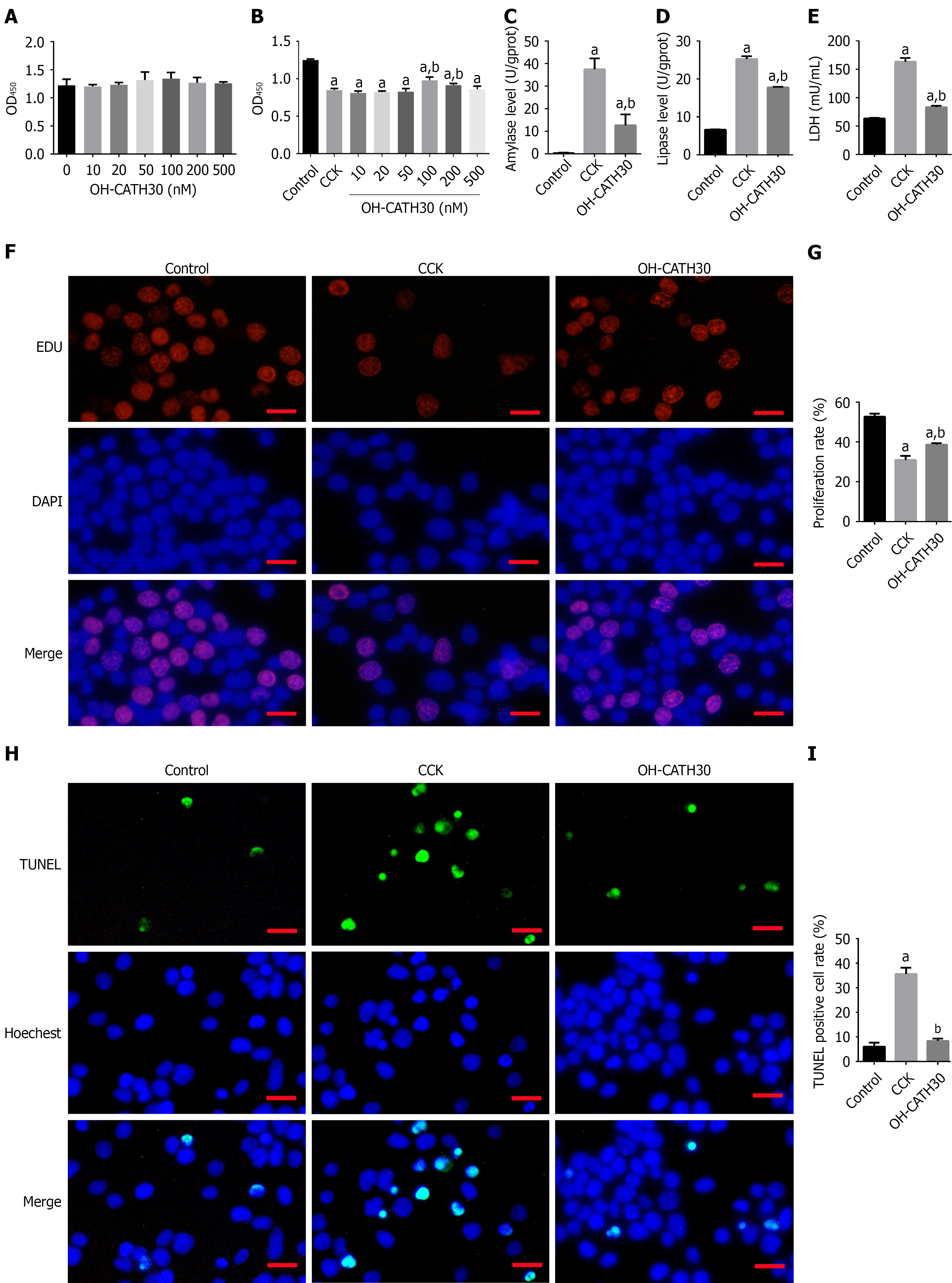
Figure 1 OH-CATH30 attenuates cholecystokinin-induced injury in pancreatic acinar cells (266-6).
A: Cell viability of 266-6 cells treated with increasing concentrations of OH-CATH30 (0-500 nM) for 24 hours, assessed using cell counting kit-8 assay; B: Cells were pretreated with 100 nM cholecystokinin (CCK) for 24 hours to induce injury, followed by treatment with OH-CATH30 at indicated concentrations. Cell viability was measured using cell counting kit-8 assay; C-E: Effects of 100 nM OH-CATH30 on cholecystokinin -injured 266-6 cells: (C) Amylase1 and (D) lipase levels in culture supernatants; (E) lactate dehydrogenase activity; F and G: Cell proliferation detected using EdU incorporation assay. Representative images (F) and quantification (G) show increased proliferation after OH-CATH30 treatment. Scale bar = 50 μm; H and I: Apoptosis detected using TUNEL staining. Representative images (H) and quantification (I) show reduced apoptosis following OH-CATH30 treatment. Scale bar = 50 μm. All data are presented as mean ± SD (n = 3 independent experiments). Statistical analysis was performed using one-way ANOVA followed by Tukey’s HSD post hoc test. aP < 0.05 vs control, bP < 0.05 vs cholecystokinin alone (0 nM OH-CATH30). CCK: Cholecystokinin; LDH: Lactate dehydrogenase.
- Citation: Li KL, Zhao Y, Shang WJ, Guo QH, Liu HT, Zhang JY, Jia XL, Liu BR. OH-CATH30 targets CD40 to suppress NF-κB signaling and promote arachidonic acid metabolic reprogramming in acute pancreatitis. World J Gastroenterol 2026; 32(36): 121753
- URL: https://www.wjgnet.com/1007-9327/full/v32/i36/121753.htm
- DOI: https://dx.doi.org/10.3748/wjg.121753