
















Copyright: ©Author(s) 2026.
World J Gastroenterol. Aug 14, 2026; 32(30): 119177
Published online Aug 14, 2026. doi: 10.3748/wjg.119177
Published online Aug 14, 2026. doi: 10.3748/wjg.119177
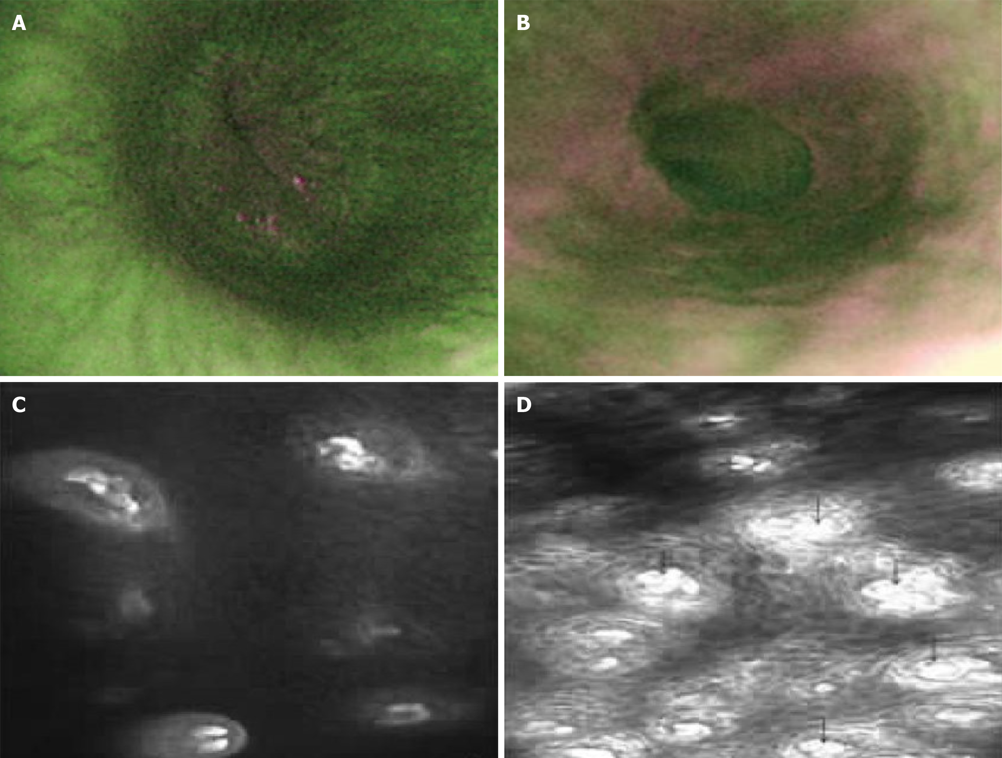
Figure 5 Autofluorescence imaging and confocal laser endomicroscopy reveal distinct features of normal esophageal mucosa and non-erosive reflux disease[27,28].
A: Normal esophageal mucosa under autofluorescence imaging; B: Longitudinal purple lines visualized under autofluorescence imaging; C: Esophageal mucosal appearance in healthy controls under confocal laser endomicroscopy; D: Dilated intercellular spaces and increased, dilated intrapapillary capillary loops (arrow) in non-erosive reflux disease patients under confocal laser endomicroscopy. Descriptions of subfigures A and B (adapted from Luo et al[27]). Citation: Luo X, Guo XX, Wang WF, Peng LH, Yang YS, Uedo N. Autofluorescence imaging endoscopy can distinguish non-erosive reflux disease from functional heartburn: A pilot study. World J Gastroenterol 2016; 22: 3845-3851. Copyright ©The Author(s) 2016. Published by Baishideng Publishing Group Inc.; descriptions of subfigures C and D (adapted from Chu et al[28]). Citation: Chu CL, Zhen YB, Lv GP, Li CQ, Li Z, Qi QQ, Gu XM, Yu T, Zhang TG, Zhou CJ, Rui-Ji, Li YQ. Microalterations of esophagus in patients with non-erosive reflux disease: In-vivo diagnosis by confocal laser endomicroscopy and its relationship with gastroesophageal reflux. Am J Gastroenterol 2012; 107: 864-874. Copyright ©The American College of Gastroenterology 2012. Published by Wolters Kluwer Health, Inc. (Supplementary material).
- Citation: Zhang WT, Xu M, Zheng Y, Wu J, Zheng YJ, Wei J, Kang T, Jiang J, Cheng Y. Endoscopic diagnosis of non-erosive reflux disease. World J Gastroenterol 2026; 32(30): 119177
- URL: https://www.wjgnet.com/1007-9327/full/v32/i30/119177.htm
- DOI: https://dx.doi.org/10.3748/wjg.119177