
















Copyright: ©Author(s) 2026.
World J Gastroenterol. Aug 14, 2026; 32(30): 119177
Published online Aug 14, 2026. doi: 10.3748/wjg.119177
Published online Aug 14, 2026. doi: 10.3748/wjg.119177
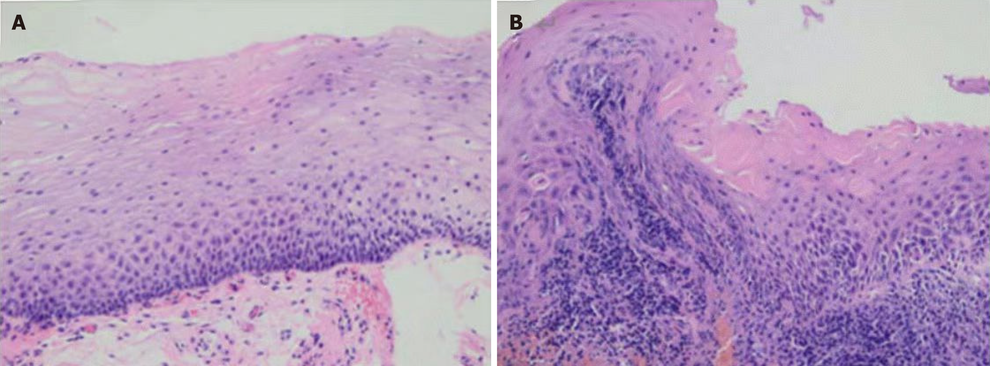
Figure 1 Histological characteristics of the esophageal epithelium stained with hematoxylin-eosin[9].
A: Mild hyperplasia of basal cells; B: Increased thickness of the basal zone and elongation of papillae in the epithelium, accompanied by neutrophil and eosinophil infiltration. Adapted from Zhang et al[9]. Citation: Zhang NN, Ma YM, Sun Q, Shi LL, Xie Y, Zou XP. Evaluation of Minimal Change Lesions Using Linked Color Imaging in Patients With Nonerosive Reflux Esophagitis. J Clin Gastroenterol 2022; 56: 405-411. Copyright ©Wolters Kluwer Health, Inc. 2021. Published by Wolters Kluwer Health, Inc. (Supplementary material).
- Citation: Zhang WT, Xu M, Zheng Y, Wu J, Zheng YJ, Wei J, Kang T, Jiang J, Cheng Y. Endoscopic diagnosis of non-erosive reflux disease. World J Gastroenterol 2026; 32(30): 119177
- URL: https://www.wjgnet.com/1007-9327/full/v32/i30/119177.htm
- DOI: https://dx.doi.org/10.3748/wjg.119177