Copyright
©The Author(s) 2025.
World J Gastroenterol. Aug 28, 2025; 31(32): 110573
Published online Aug 28, 2025. doi: 10.3748/wjg.v31.i32.110573
Published online Aug 28, 2025. doi: 10.3748/wjg.v31.i32.110573
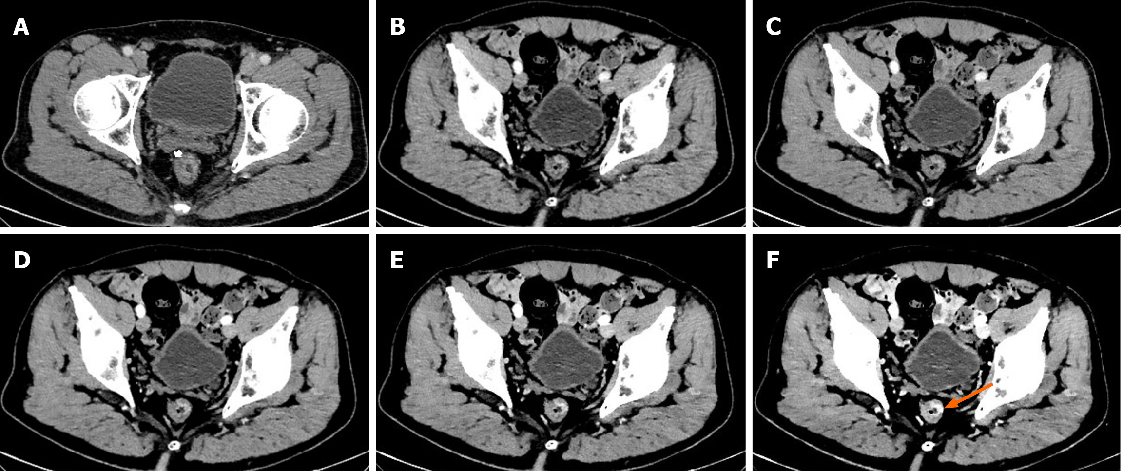
Figure 4 T-staging in axial polyenergetic image, oblique axial polyenergetic image and 40-70 keV oblique axial virtual monoenergetic images for T2 stage by pathology.
A middle rectal cancer patient with axial polyenergetic image (PEI), oblique PEI and 40-70 keV oblique axial (OA) virtual monoenergetic images (VMIs), was proved to be T2 by pathology. A: Axial PEI showed a smooth outer border of the thickened rectal wall with peritumoral fat stranding (PFS; white arrow), suggesting stage T3; B-F: Oblique PEI (B) and 70-40 keV OA VMIs (C-F) demonstrated the lesion located in the left anterior wall (orange arrow) of the rectal wall and the outer border of the thickened rectal wall was smooth. No spiculations extending into the perirectal fat and PFS was found, suggesting stage T2. Compared to axial PEI (A), the lesion was better visualized at 40-50 keV VMIs due to increased lesion attenuation and hyper-enhancement. Among all images, the 40-50 keV OA VMIs (E and F) performed best.
- Citation: Chen FX, Jiang KK, Zhu JF, Wang MR, Fan XL, Yang JS, He BS. Diagnostic accuracy of dual-layer spectral computed tomography virtual monoenergetic imaging with multiplanar reformation for T-staging of colorectal cancer. World J Gastroenterol 2025; 31(32): 110573
- URL: https://www.wjgnet.com/1007-9327/full/v31/i32/110573.htm
- DOI: https://dx.doi.org/10.3748/wjg.v31.i32.110573


