Copyright
©The Author(s) 2025.
World J Gastroenterol. Aug 21, 2025; 31(31): 109994
Published online Aug 21, 2025. doi: 10.3748/wjg.v31.i31.109994
Published online Aug 21, 2025. doi: 10.3748/wjg.v31.i31.109994
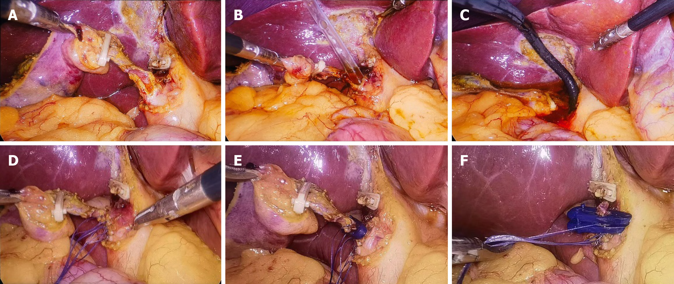
Figure 2 Surgical procedure.
A: Clip the cystic artery and the gallbladder-side of the cystic duct, make a transverse incision on the cystic duct 1-2 cm from the common bile duct, and extend a longitudinal incision from the transverse incision along the cystic duct to its junction with the common bile duct; B: Sequentially insert 6F, 8F, 10F, 12F, 14F, and 16F dilators through the incision to dilate the bile duct; C: Insert a choledochoscope through the cystic duct to explore the common bile duct and remove stones; D: Perform interrupted suture at the junction of the cystic duct and common bile duct with absorbable sutures; E: Clip the cystic duct with absorbable clips; F: Complete cholecystectomy.
- Citation: Zhu DS, Zhang Z, Huang XR, Zhang JZ, Zhang ZW, Guo XY, Zheng H, Guo T, Yu YH. Textbook outcome and associated risk factors in laparoscopic transcystic common bile duct exploration. World J Gastroenterol 2025; 31(31): 109994
- URL: https://www.wjgnet.com/1007-9327/full/v31/i31/109994.htm
- DOI: https://dx.doi.org/10.3748/wjg.v31.i31.109994


