Copyright
©The Author(s) 2025.
World J Gastroenterol. Jul 28, 2025; 31(28): 108990
Published online Jul 28, 2025. doi: 10.3748/wjg.v31.i28.108990
Published online Jul 28, 2025. doi: 10.3748/wjg.v31.i28.108990
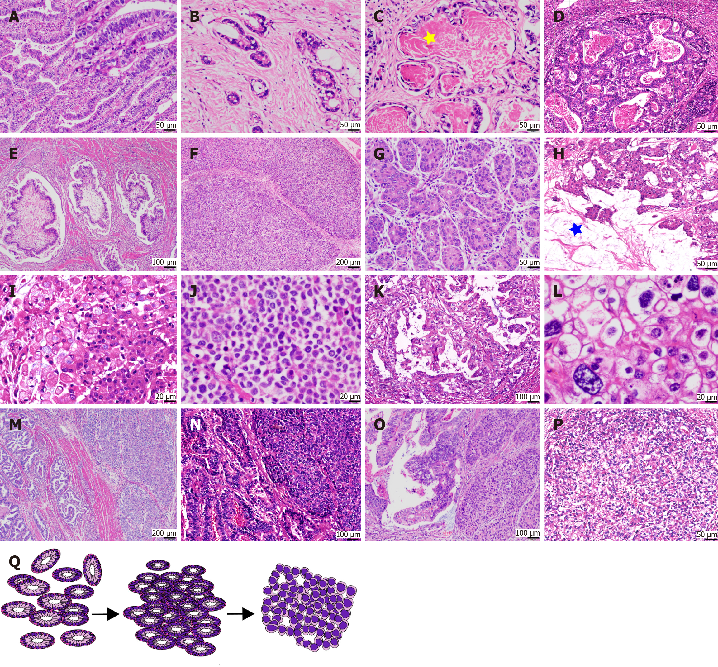
Figure 2 Histopathological diversity of gastric adenocarcinoma with primitive enterocyte phenotype.
A and B: Tubular-papillary subtype of gastric adenocarcinoma with primitive enterocyte phenotype characterized by glandular structures with supranuclear and subnuclear vacuoles, creating a distinctive “piano keyboard-like” architecture; C: Luminal eosinophilic secretions within glandular spaces (asterisks); D and E: Uncommon architectural patterns, including sieve-like/sac-like formations and villous projections with tumor cells exhibiting flattened morphology; F and G: Solid subtype resembling hepatocellular carcinoma, featuring large polygonal tumor cells with eosinophilic or clear cytoplasm arranged in nested sheets; H and I: Intracellular and extracellular mucin deposition (asterisks) observed in select cases; J: Rare poorly cohesive growth pattern akin to gastric poorly cohesive carcinoma; K and L: High-grade cytological atypia, including nuclear pleomorphism and prominent nucleoli; M-O: Transitional zones demonstrating admixture and migratory progression between hepatoid adenocarcinoma and gastric adenocarcinoma with enteroblastic differentiation components; P: Predominance of clear cytoplasmic features over eosinophilic differentiation in solid areas; Q: Schematic representation of phenotypic evolution from tubular-papillary to solid morphology.
- Citation: Li HQ, Zheng LQ, Huang WT, Yu XB, Zhang X, Lin L, Lv SS, Yan XY, Chen XY. Clinicopathological significance of histological diversity in gastric adenocarcinoma with primitive enterocyte phenotype: A methylation-driven aggressive entity. World J Gastroenterol 2025; 31(28): 108990
- URL: https://www.wjgnet.com/1007-9327/full/v31/i28/108990.htm
- DOI: https://dx.doi.org/10.3748/wjg.v31.i28.108990


