Copyright
©The Author(s) 2025.
World J Gastroenterol. Jul 21, 2025; 31(27): 106166
Published online Jul 21, 2025. doi: 10.3748/wjg.v31.i27.106166
Published online Jul 21, 2025. doi: 10.3748/wjg.v31.i27.106166
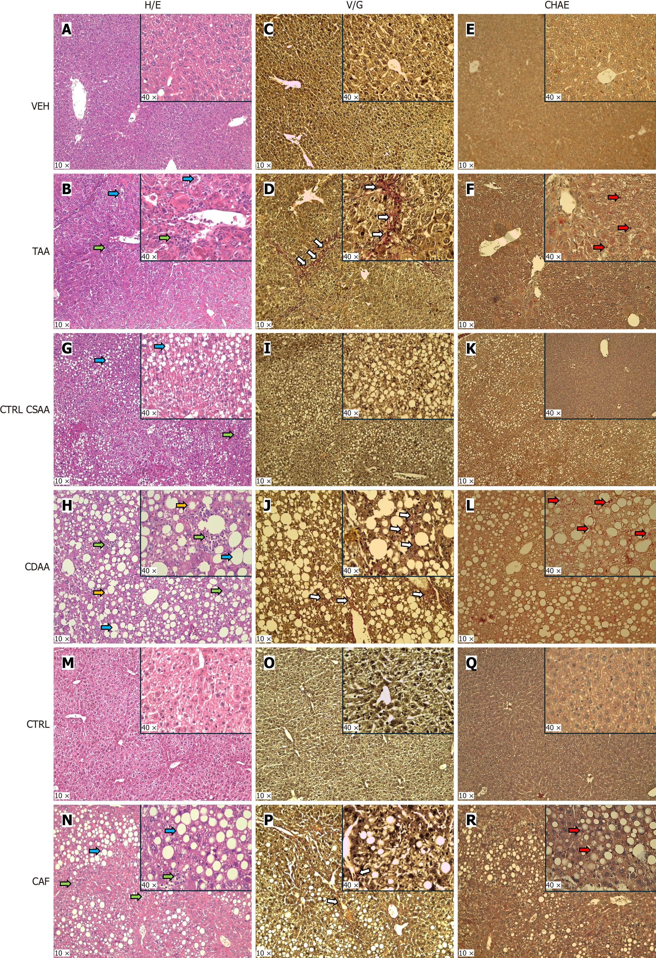
Figure 3 Representative pictures of liver histology.
A: Hematoxylin and eosin staining (H/E) in the liver of vehicle-injected mouse; B: H/E in liver of thioacetamide (TAA)-injected mouse; C: Van Gieson’s staining (VG) in the liver of vehicle-injected mouse; D: VG in the liver of TAA-injected mouse; E: Naphthol-AS-D-chloroacetate esterase staining (CHAE) in the liver of vehicle-injected mouse; F: CHAE in the liver of TAA-injected mouse; G: H/E in the liver of choline-sufficient L-amino acid-defined control (CTRL CSAA) diet-fed mouse; H: H/E in the liver of choline-deficient L-amino acid-defined (CDAA) diet-fed mouse; I: VG in the liver of CTRL CSAA diet-fed mouse; J: VG in the liver of CDAA diet-fed mouse; K: CHAE in the liver of CTRL CSAA diet-fed mouse L: CHAE in the liver of CDAA diet-fed mouse; M: H/E in the liver of control (CTRL) diet-fed mouse; N: H/E in the liver of cafeteria (CAF) diet-fed mouse; O: VG in the liver of CTRL diet-fed mouse; P: VG in the liver of CAF diet-fed mouse; Q: CHAE in the liver of CTRL diet-fed mouse; R: CHAE in the liver of CAF diet-fed mouse. The blue arrow points to the lobular inflammation. The green arrow points to the steatotic hepatocytes. The orange arrow points to the ballooning of hepatocytes. The white arrow points to the fibrotic tissue. The red arrow points to the neutrophils that infiltrate the liver. H/E: Hematoxylin and eosin staining; TAA: Thioacetamide; VG: Van Gieson’s staining; CHAE: Chloroacetate esterase staining; CTRL CSAA: Choline-sufficient L-amino acid-defined control; CDAA: Choline-deficient L-amino acid-defined; CAF: Cafeteria.
- Citation: Feješ A, Belvončíková P, Bečka E, Strečanský T, Pastorek M, Janko J, Filová B, Babál P, Šebeková K, Borbélyová V, Gardlík R. Myeloperoxidase, extracellular DNA and neutrophil extracellular trap formation in the animal models of metabolic dysfunction-associated steatotic liver disease. World J Gastroenterol 2025; 31(27): 106166
- URL: https://www.wjgnet.com/1007-9327/full/v31/i27/106166.htm
- DOI: https://dx.doi.org/10.3748/wjg.v31.i27.106166


