Copyright
©The Author(s) 2025.
World J Gastroenterol. Jul 14, 2025; 31(26): 109500
Published online Jul 14, 2025. doi: 10.3748/wjg.v31.i26.109500
Published online Jul 14, 2025. doi: 10.3748/wjg.v31.i26.109500
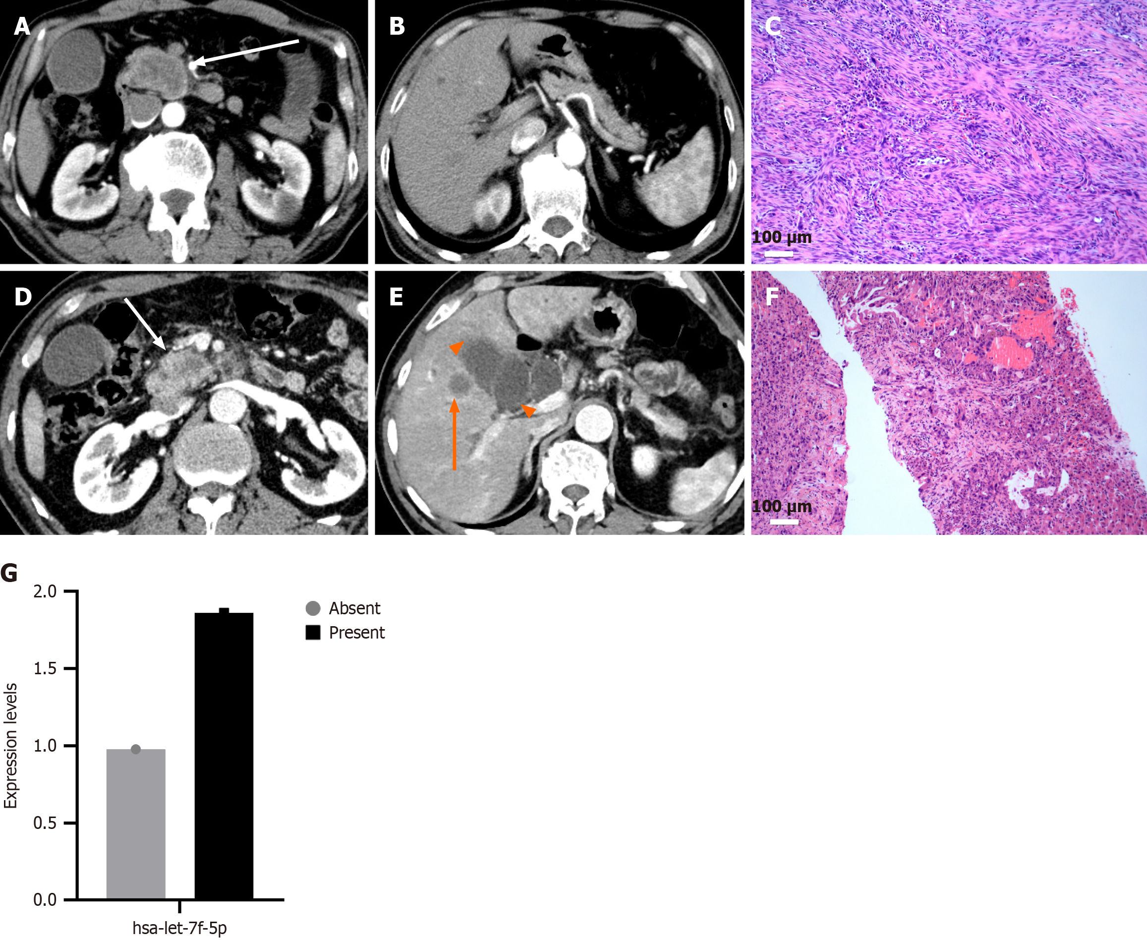
Figure 5 Representative computed tomography and pathological findings in patients with pancreatic cancer.
A and B: A 64-year-old male with pancreatic head cancer (white arrow), lesion size approximately 4.38 cm in diameter, showing no evidence of vascular invasion or lymph node metastasis; C: Pathological section at 100 × magnification shows tumor cells scattered singly within fibrous connective tissue. TNM stage: T3N0M0 (stage IIA); D and E: A 68-year-old male with pancreatic head cancer (white arrow), lesion size approximately 4.0 cm in diameter, with a ring-enhancing metastatic lesion in the right liver lobe (orange arrow) and an enlarged gallbladder (orange arrowhead); F: Pathological section at 100 × magnification shows metastatic adenocarcinoma within trabecular liver tissue, characterized by atypical glandular structures and scattered tumor cells. TNM stage: T2NxM1 (stage IV); G: Serum exosomal hsa-let-7f-5p expression levels in patients with localized pancreatic cancer and those with metastasis were 09768 and 1.8632, respectively.
- Citation: Ren S, Song LN, Zhao R, Tian Y, Wang ZQ. Serum exosomal hsa-let-7f-5p: A potential diagnostic biomarker for metastatic pancreatic cancer detection. World J Gastroenterol 2025; 31(26): 109500
- URL: https://www.wjgnet.com/1007-9327/full/v31/i26/109500.htm
- DOI: https://dx.doi.org/10.3748/wjg.v31.i26.109500


