Copyright
©The Author(s) 2025.
World J Gastroenterol. Jul 14, 2025; 31(26): 107044
Published online Jul 14, 2025. doi: 10.3748/wjg.v31.i26.107044
Published online Jul 14, 2025. doi: 10.3748/wjg.v31.i26.107044
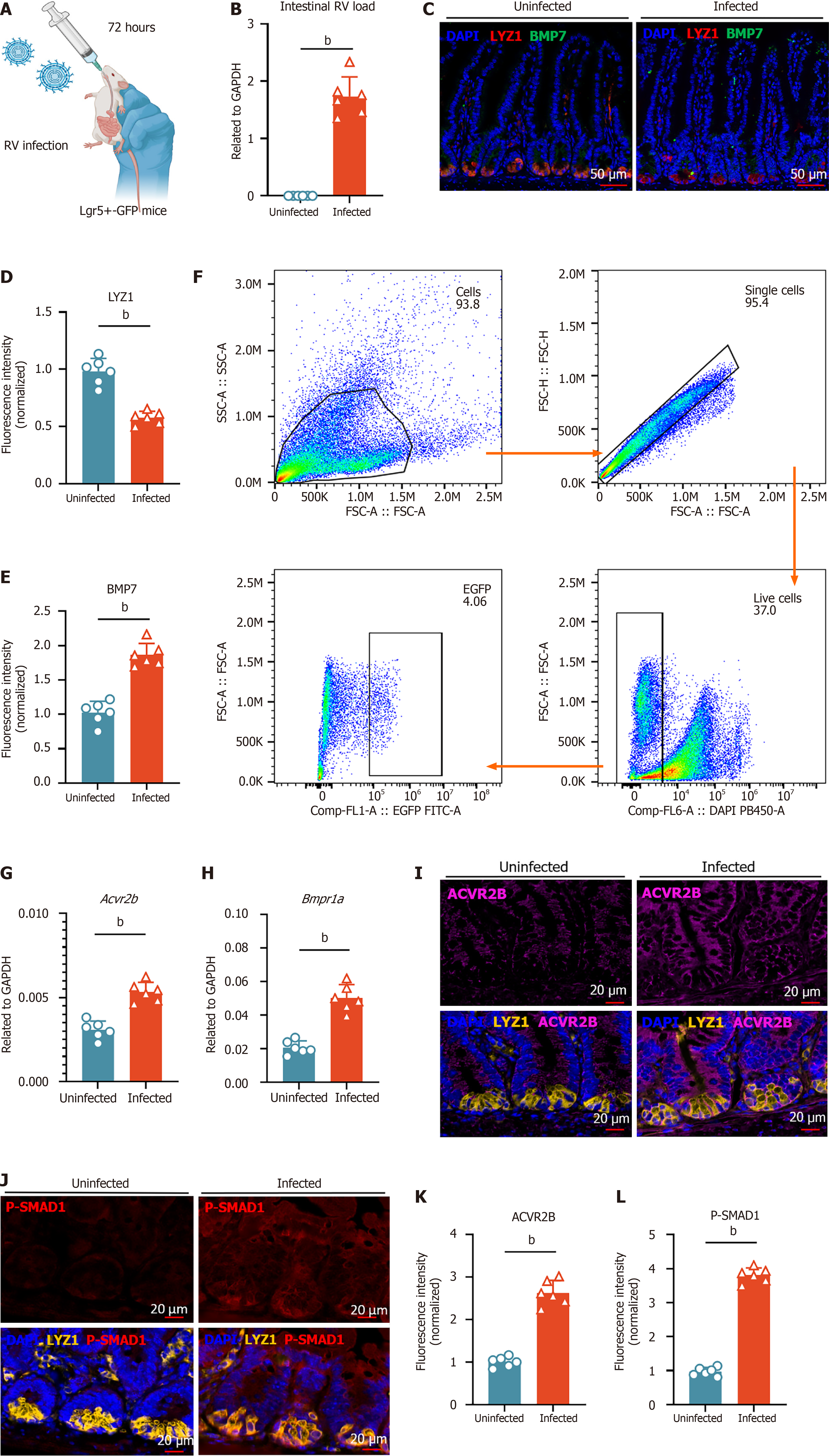
Figure 5 Functional validation of Paneth-intestinal stem cells bone morphogenic protein 7 pathway upregulation under rotavirus infection.
A: Leucine-rich repeat-containing G protein-coupled receptor 5+(Lgr5+)-green fluorescent protein mice aged 8 weeks were orally gavaged with the rotavirus enteric cytopathogenic human orphan virus strain and sacrificed 72 hours post-infection; B: Quantification of intestinal rotavirus load in uninfected and infected mice (n = 6, mean ± SD), measured by reverse transcription quantitative polymerase chain reaction and normalized to glyceraldehyde-3-phosphate dehydrogenase; C: Immunofluorescence (IF) staining of bone morphogenic protein 7 (BMP7) (green) and Paneth cell marker lysozyme 1 (LYZ1) (red) in uninfected and infected mice. Nuclei were counterstained with 4’,6-diamidino-2-phenylindole (DAPI) (blue). Scale bar = 50 μm; D and E: Quantification of LYZ1 and BMP7 fluorescence intensity in stem cells from uninfected and infected mice. Fluorescence intensity was normalized to the uninfected group (set to 1); F: Flow cytometric analysis for isolating Lgr5+ intestinal stem cells from uninfected and infected mice; G and H: Activin A receptor type 2B (Acvr2b) and BMP receptor type 1A expression in sorted Lgr5+ intestinal stem cells from uninfected and infected mice (n = 6, mean ± SD), measured by quantitative real-time polymerase chain reaction and normalized to glyceraldehyde-3-phosphate dehydrogenase; I: IF staining of ACVR2B (magenta) in uninfected and infected mice. Bottom panels show merged images of ACVR2B (magenta) with Paneth cell marker LYZ1 (yellow) and DAPI (blue). Scale bar = 20 μm; J: IF staining of phosphorylation of SMAD1 (red) in uninfected and infected mice. Right panels show merged images of phosphorylation of SMAD1 (red) with LYZ1 (yellow) and DAPI (blue). Scale bar = 20 μm; K and L: Quantification of ACVR2B and phosphorylation of Smad1 fluorescence intensity in stem cells from uninfected and infected mice. Fluorescence intensity was normalized to the uninfected group (set to 1). Statistical significance was determined using the non-parametric Mann-Whitney test between groups. bP < 0.01. RV: Rotavirus; Lgr5+: Leucine-rich repeat-containing G protein-coupled receptor 5+; GFP: Green fluorescent protein; GAPDH: Glyceraldehyde-3-phosphate dehydrogenase; DAPI: 4’,6-diamidino-2-phenylindole; Lyz: Lysozyme; BMP: Bone morphogenic protein; Acvr2b: Activin A receptor type 2B; Bmpr1a: Bone morphogenic protein receptor type 1A; P-SMAD1: Phosphorylation of SMAD1.
- Citation: Bu XY, Tan HY, Wang AM, Wei MT, Pan S, Gao JZ, Li YH, Qian GX, Chen ZH, Ye C, Jia WD. Paneth cells inhibit intestinal stem cell proliferation through the bone morphogenic protein 7 pathway under rotavirus-mediated intestinal injury. World J Gastroenterol 2025; 31(26): 107044
- URL: https://www.wjgnet.com/1007-9327/full/v31/i26/107044.htm
- DOI: https://dx.doi.org/10.3748/wjg.v31.i26.107044


