Copyright
©The Author(s) 2025.
World J Gastroenterol. Jun 28, 2025; 31(24): 108234
Published online Jun 28, 2025. doi: 10.3748/wjg.v31.i24.108234
Published online Jun 28, 2025. doi: 10.3748/wjg.v31.i24.108234
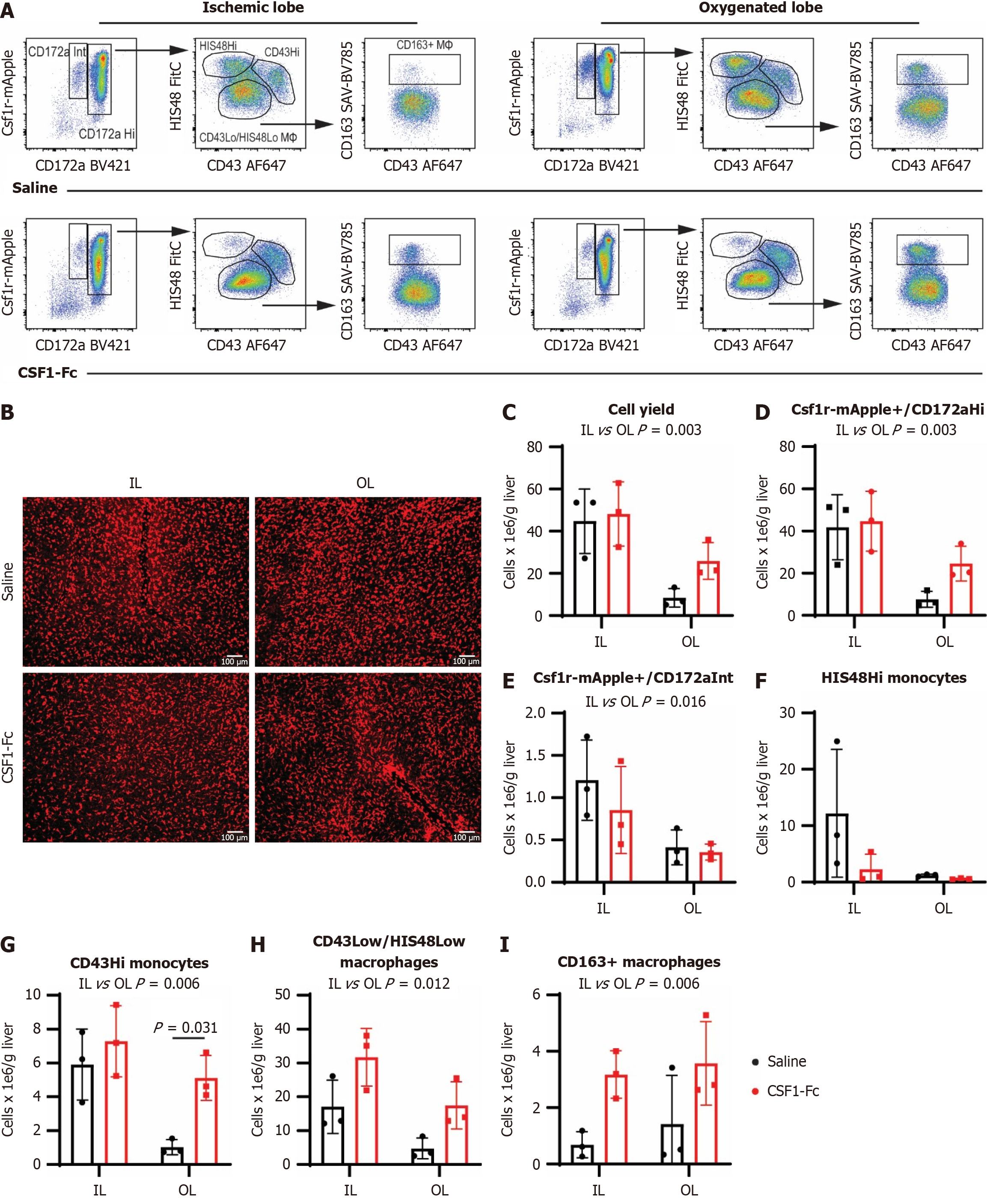
Figure 5 Colony stimulating factor 1-Fc mobilises non-classical monocytes into the liver and increases CD163+ macrophages.
Non-parenchymal cells were isolated from disaggregated livers harvested from Csf1r-mApple rats 48 hours post ischemia-reperfusion (I/R). A: Csf1r-mApple expression and CD172a (SIRPa) were used to gate myeloid cells, and further subdivided into CH172Hi (monocytes and macrophages) and CD172Intermediate (neutrophils); B: Representative whole-mount imaging of fresh unfixed tissues from Csf1r-mApple transgenic rats 48 hours post I/R using a spinning disc confocal microscope, n = 3 for all groups and results were analysed with ordinary two-way ANOVA with Tukey’s multiple comparisons for B-H; C-I: CD43 low/HIS48 low cells were gated as macrophages, HIS48Hi as classical monocytes and CD43Hi as non-classical monocytes. The liver macrophage marker CD163 was expressed on a subset of CD43 low/HIS48 low cells and the impact of colony stimulating factor 1 treatment on cell yield and the different subpopulations in the ischemic and oxygenated lobes were quantified; Data show mean and standard deviation. IL: Ischemic lobe; OL: Oxygenated lobe; CSF: Colony stimulating factor.
- Citation: Schulze S, Keshvari S, Miller GC, Bridle KR, Hume DA, Irvine KM. Perisurgical colony stimulating factor one treatment ameliorates liver ischaemia/reperfusion injury in rats. World J Gastroenterol 2025; 31(24): 108234
- URL: https://www.wjgnet.com/1007-9327/full/v31/i24/108234.htm
- DOI: https://dx.doi.org/10.3748/wjg.v31.i24.108234


