Copyright
©The Author(s) 2025.
World J Gastroenterol. Jun 21, 2025; 31(23): 106261
Published online Jun 21, 2025. doi: 10.3748/wjg.v31.i23.106261
Published online Jun 21, 2025. doi: 10.3748/wjg.v31.i23.106261
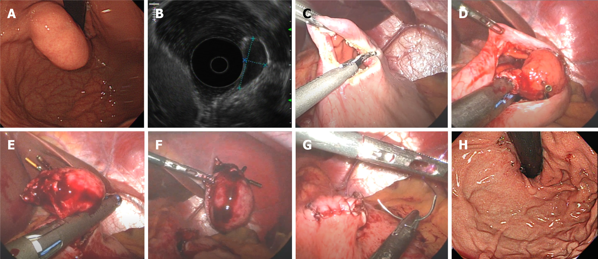
Figure 3 Laparoscopic wedge resection.
A: Endoscopic view of the submucosal tumor in the cardia; B: Endoscopic ultrasound image showing a tumor originating from the muscularis propria; C and D: Dissection of the anterior wall of the stomach to obtain an intragastric view with the submucosal tumor marked with clips; E and F: A laparoscopic linear stapler was used to perform wedge resection; G: Surgical suturing of the stomach; H: Endoscopic follow-up 6 months later showing no deformities.
- Citation: Lee AY, Lim SG, Cho JY, Kim S, Lee KM, Shin SJ, Noh CK, Lee GH, Hur H, Han SU, Son SY, Song JH. Comparison of treatment strategies for submucosal tumors originating from the muscularis propria at esophagogastric junction or cardia. World J Gastroenterol 2025; 31(23): 106261
- URL: https://www.wjgnet.com/1007-9327/full/v31/i23/106261.htm
- DOI: https://dx.doi.org/10.3748/wjg.v31.i23.106261


