Copyright
©The Author(s) 2025.
World J Gastroenterol. Jun 21, 2025; 31(23): 106261
Published online Jun 21, 2025. doi: 10.3748/wjg.v31.i23.106261
Published online Jun 21, 2025. doi: 10.3748/wjg.v31.i23.106261
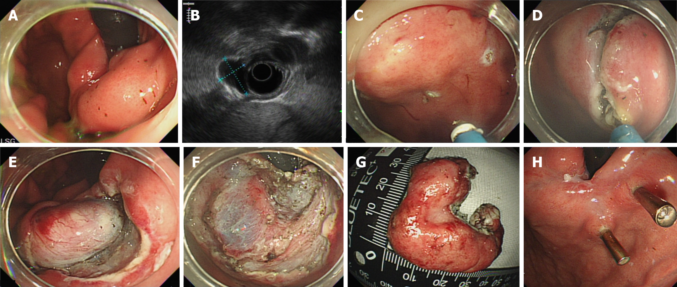
Figure 1 Endoscopic submucosal dissection.
A: Endoscopic view of a submucosal tumor in the cardia of the stomach; B: Endoscopic ultrasonography showing a hypoechoic submucosal tumor in the muscularis propria; C: Circumferential markings around the lesion; D and E: Mucosal incision along the marked points after submucosal injection; F: The tumor was completely resected macroscopically; G: Resected specimen; H: Endoscopic view at the 3-month follow-up.
- Citation: Lee AY, Lim SG, Cho JY, Kim S, Lee KM, Shin SJ, Noh CK, Lee GH, Hur H, Han SU, Son SY, Song JH. Comparison of treatment strategies for submucosal tumors originating from the muscularis propria at esophagogastric junction or cardia. World J Gastroenterol 2025; 31(23): 106261
- URL: https://www.wjgnet.com/1007-9327/full/v31/i23/106261.htm
- DOI: https://dx.doi.org/10.3748/wjg.v31.i23.106261


