Copyright
©The Author(s) 2025.
World J Gastroenterol. Jun 21, 2025; 31(23): 103848
Published online Jun 21, 2025. doi: 10.3748/wjg.v31.i23.103848
Published online Jun 21, 2025. doi: 10.3748/wjg.v31.i23.103848
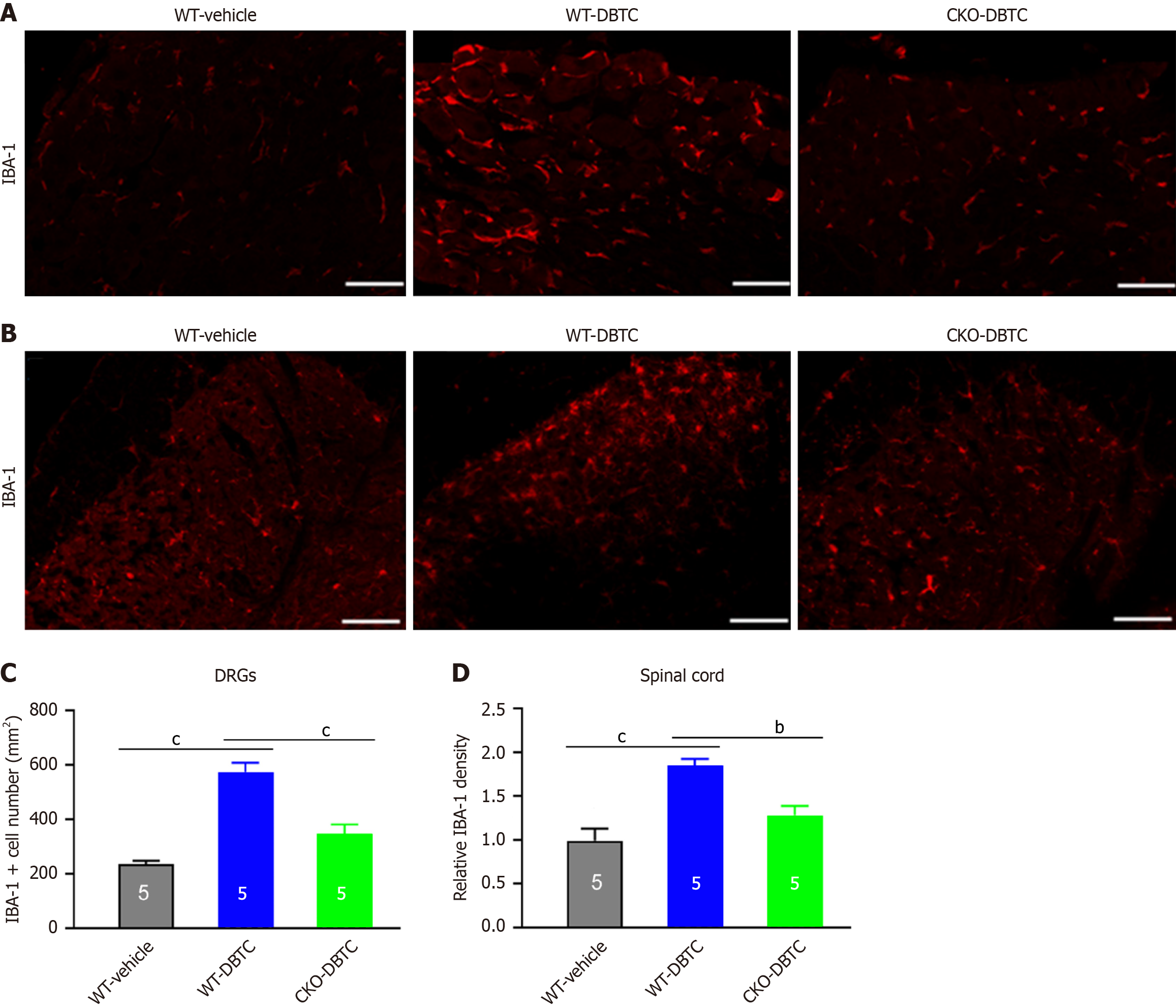
Figure 4 Increased macrophage infiltration into the DRG and microglial activation in the spinal cord of mice with chronic pancreatitis.
A and B: Representative images of immunofluorescence staining of ionized calcium-binding adapter molecule 1 (IBA-1) on the T8-12 dorsal root ganglion neurons (A) and spinal cord (B); C: The number of IBA-1-positive cells; D: Relative IBA-1-positive density (normalized to WT-vehicle group) in the dorsal spinal cord was analyzed for each group of mice. Scale bar, 50 μm (A) and 100 μm (B). n = 5 mice. Data are shown as mean ± SEM. bP < 0.01, cP < 0.001, statistical comparison were conducted with one-way analysis of variance with Sidak’s post hoc test. WT: Wild type; CKO: Conditional knockout; DBTC: Dibutyltin dichloride; DRG: Dorsal root ganglion; IBA-1: Ionized calcium binding adaptor molecule 1.
- Citation: Wang B, Ge JY, Wu JN, Xu JH, Cao XH, Chang N, Zhou X, Jing PB, Liu XJ, Wu Y. Endothelin A receptor in nociceptors is essential for persistent mechanical pain in a chronic pancreatitis of mouse model. World J Gastroenterol 2025; 31(23): 103848
- URL: https://www.wjgnet.com/1007-9327/full/v31/i23/103848.htm
- DOI: https://dx.doi.org/10.3748/wjg.v31.i23.103848


