Copyright
©The Author(s) 2025.
World J Gastroenterol. Jun 7, 2025; 31(21): 107601
Published online Jun 7, 2025. doi: 10.3748/wjg.v31.i21.107601
Published online Jun 7, 2025. doi: 10.3748/wjg.v31.i21.107601
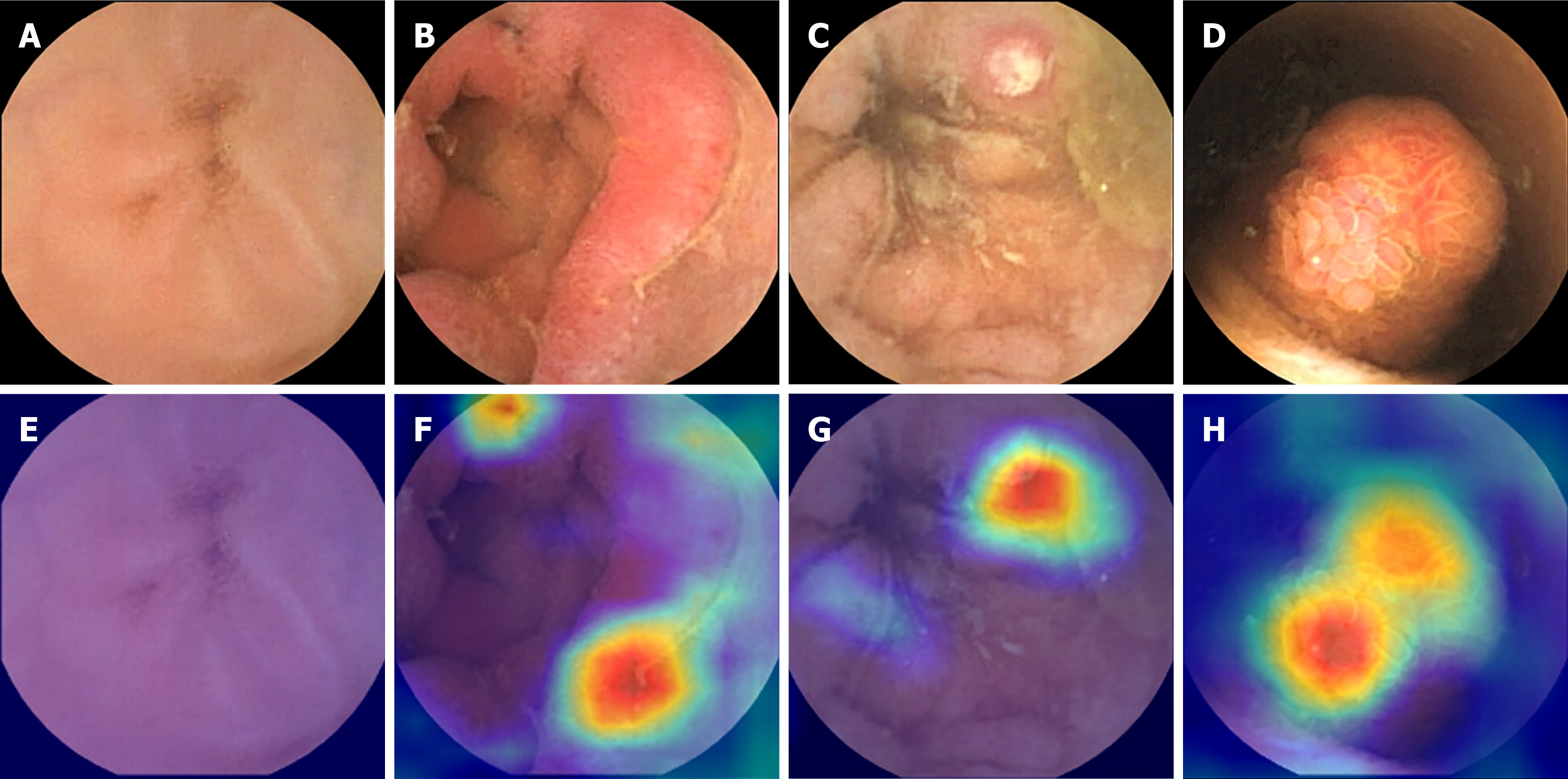
Figure 2 Lesion detection by applying gradient-weighted class activation mapping on representative video capsule endoscopy images, including normal mucosa, erosions/erythema, ulcer, and polyps images.
A-D: White-light imaging capsule endoscopic image; E-H: Gradient-weighted class activation mapping image. A and E: Normal mucosa; B and F: Erosions/erythema; C and G: Ulcer; D and H: Polyps. Grad-CAM: Gradient-weighted class activation mapping; VCE: Video capsule endoscopy.
- Citation: Huang YH, Lin Q, Jin XY, Chou CY, Wei JJ, Xing J, Guo HM, Liu ZF, Lu Y. Classification of pediatric video capsule endoscopy images for small bowel abnormalities using deep learning models. World J Gastroenterol 2025; 31(21): 107601
- URL: https://www.wjgnet.com/1007-9327/full/v31/i21/107601.htm
- DOI: https://dx.doi.org/10.3748/wjg.v31.i21.107601


