Copyright
©The Author(s) 2025.
World J Gastroenterol. May 28, 2025; 31(20): 100192
Published online May 28, 2025. doi: 10.3748/wjg.v31.i20.100192
Published online May 28, 2025. doi: 10.3748/wjg.v31.i20.100192
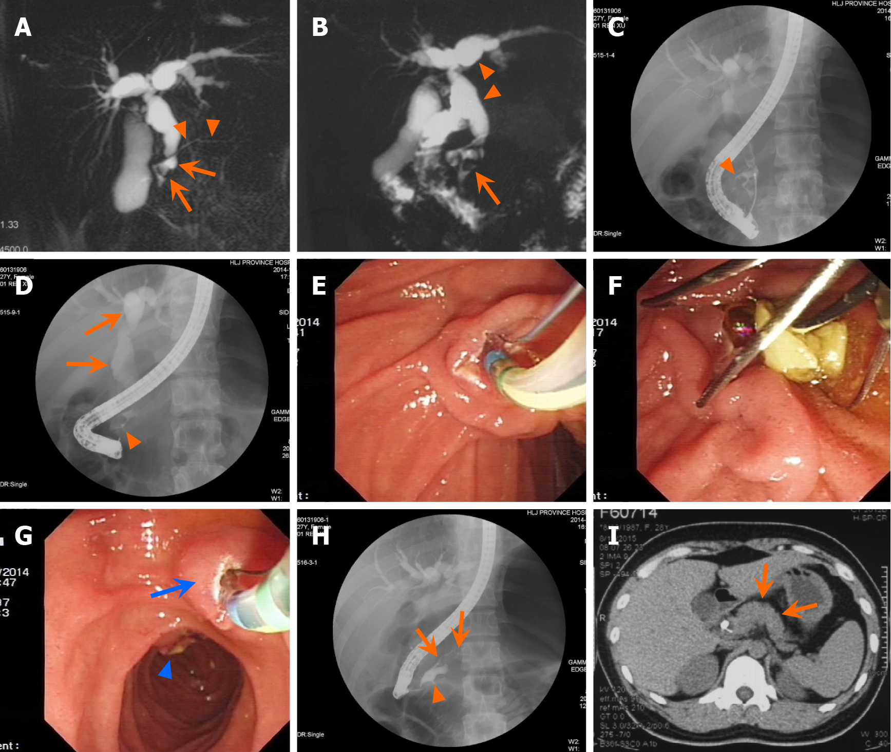
Figure 4 A case of complex pancreaticobiliary maljunction with complete pancreatic divisum and accompanied with stones in both the dorsal duct and the common bile duct.
A: Magnetic resonance cholangiopancreatography (MRCP) shows notable localized dilation of the dorsal duct (arrow) and a pancreatic stone (arrow), accompanied by a markedly narrowed upstream dorsal duct (arrowhead) and dilation of the intrahepatic and extrahepatic bile ducts. The ventral duct is not visualized; B: MRCP displays the presence of a common bile duct stone (arrow) and dilated dorsal duct, along with dilation of the intrahepatic and extrahepatic bile ducts (arrowhead); C: Cholangiography via the major papilla shows localized dilation of the dorsal duct and a pancreatic stone (arrowhead), indicating an anomalous junction between the dorsal duct and common bile duct; D: Cholangiography via the major papilla shows a large stone in the dorsal duct (8 mm × 12 mm; arrowhead), along with dilation of the intrahepatic and extrahepatic bile ducts progressing towards a choledochal cyst (arrow); E: Endoscopic sphincterotomy is being performed; F: The common bile duct stone is extracted using a basket; G: Endoscopic minor papillotomy (arrow) is being carried out and the major papilla can be seen in distance; H: Pancreatography via the minor papilla after removed the pancreatic stone reveals localized dilation of the dorsal duct (arrowhead) and a narrowed upstream dorsal duct (arrow); I: Computed tomography scan indicates no agenesis of the dorsal pancreas (i.e. absence of the pancreatic body and tail (arrow).
- Citation: Ren X, Qu YP, Xia T, Tang XF. Technical success, clinical efficacy, and safety of endoscopic minor papilla interventions for symptomatic pancreatic diseases. World J Gastroenterol 2025; 31(20): 100192
- URL: https://www.wjgnet.com/1007-9327/full/v31/i20/100192.htm
- DOI: https://dx.doi.org/10.3748/wjg.v31.i20.100192


