Copyright
©The Author(s) 2025.
World J Gastroenterol. May 21, 2025; 31(19): 106814
Published online May 21, 2025. doi: 10.3748/wjg.v31.i19.106814
Published online May 21, 2025. doi: 10.3748/wjg.v31.i19.106814
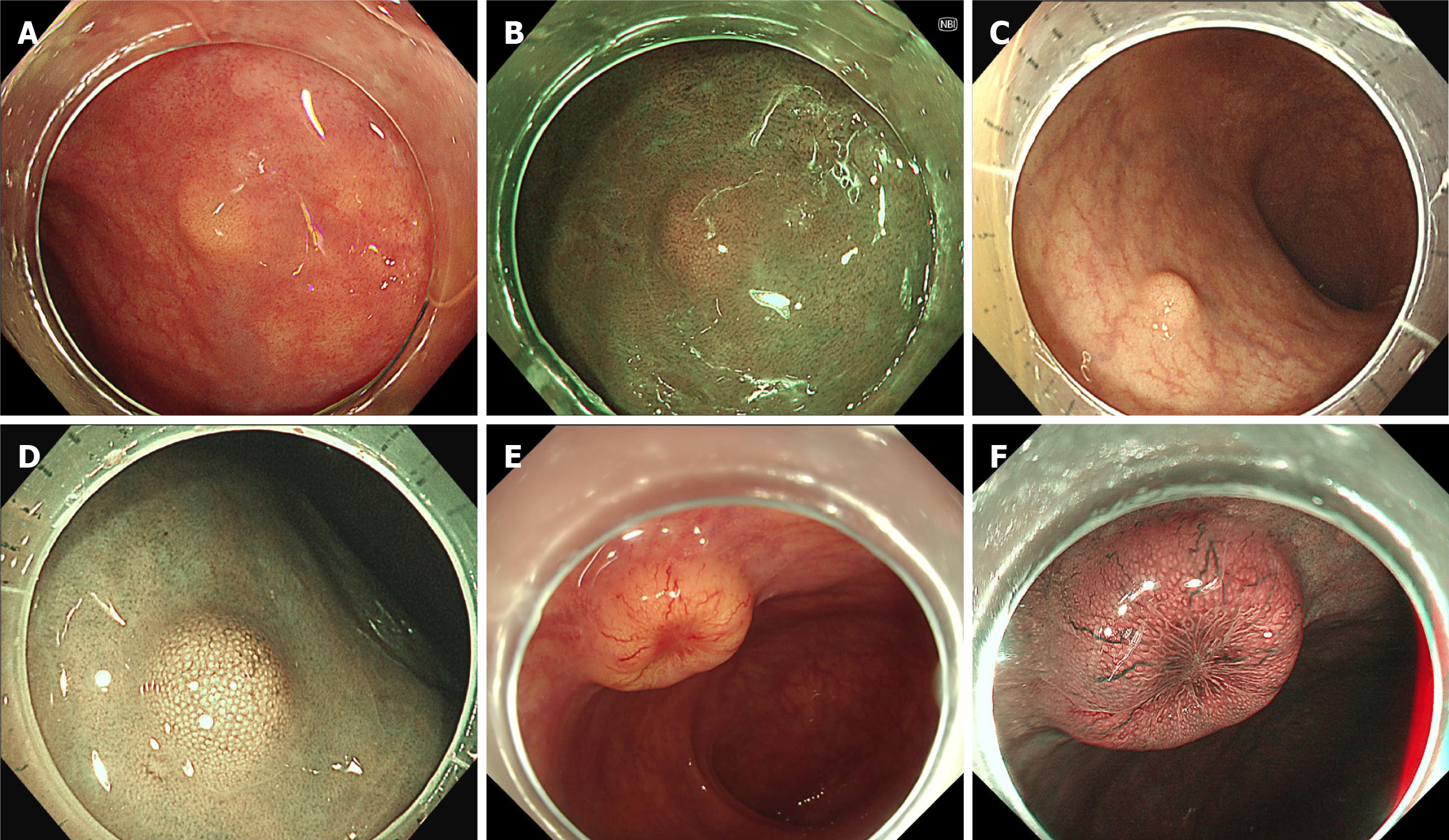
Figure 1 Endoscopic diagnosis of rectal neuroendocrine tumors.
A and B: The rectal neuroendocrine tumor typically appears as a small, yellowish, and subepithelial tumor-like lesion without surface change under white light or narrow band imaging as shown in case 1; C and D: For lesions with more significant elevation, there may be enlarged surface pit openings, but the overall shape remains unchanged, as seen in case 2; E and F: As the lesion grows larger, central depression may develop, accompanied by dilated micro-vessels, as illustrated in case 3.
- Citation: Liu JN, Chen H, Fang N. Current status of endoscopic resection for small rectal neuroendocrine tumors. World J Gastroenterol 2025; 31(19): 106814
- URL: https://www.wjgnet.com/1007-9327/full/v31/i19/106814.htm
- DOI: https://dx.doi.org/10.3748/wjg.v31.i19.106814


