Copyright
©The Author(s) 2025.
World J Gastroenterol. May 21, 2025; 31(19): 104897
Published online May 21, 2025. doi: 10.3748/wjg.v31.i19.104897
Published online May 21, 2025. doi: 10.3748/wjg.v31.i19.104897
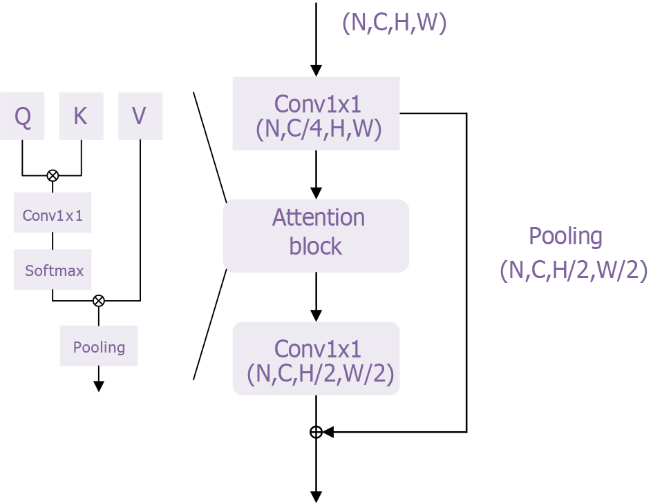
Figure 4 Transformer module of the transformer model.
This figure provides an intuitive visualization of the core structure and functionality of the transformer architecture. The self-attention mechanism and multi-head attention mechanism effectively capture and process relationships among different features in the input data. This structure enhances the model’s ability to comprehend contextual information while enabling parallel processing, which improves computational efficiency. By presenting a visual representation, researchers and clinicians can better understand how the transformer model functions in esophageal adenocarcinoma classification, increasing confidence in the model’s reliability. Furthermore, this figure supports the study’s findings by offering clear structural explanations and visual validations, demonstrating the effectiveness and robustness of the transformer model in tackling complex medical problems. This further underscores its potential for clinical applications.
- Citation: Wei W, Zhang XL, Wang HZ, Wang LL, Wen JL, Han X, Liu Q. Application of deep learning models in the pathological classification and staging of esophageal cancer: A focus on Wave-Vision Transformer. World J Gastroenterol 2025; 31(19): 104897
- URL: https://www.wjgnet.com/1007-9327/full/v31/i19/104897.htm
- DOI: https://dx.doi.org/10.3748/wjg.v31.i19.104897


